[English] 日本語
Yorodumi
- PDB-1urj: Single stranded DNA-binding protein(ICP8) from Herpes simplex virus-1 -

+
Open data


ID or keywords:

Loading...

-
Basic information

Entry
Database: PDB / ID: 1urj
TitleSingle stranded DNA-binding protein(ICP8) from Herpes simplex virus-1
ComponentsMAJOR DNA-BINDING PROTEIN
KeywordsDNA BINDING PROTEIN / DNA-BINDING PROTEIN / SSB / ICP8 / HSV-1 / DNA-BINDING / DNA REPLICATION / ZINC-FINGER / NUCLEAR PROTEIN.
Function / homology
Function and homology information


nuclear viral factory / bidirectional double-stranded viral DNA replication / single-stranded DNA binding / DNA replication / host cell nucleus / DNA binding / metal ion binding
Similarity search - Function
DNA polymerase; domain 1 - #560 / Viral ssDNA binding protein, head domain / Viral ssDNA-binding protein / DBP-like superfamily / Viral ssDNA binding protein, head domain / ssDNA binding protein / Delta-Endotoxin; domain 1 / DNA polymerase; domain 1 / Up-down Bundle / Orthogonal Bundle / Mainly Alpha
Similarity search - Domain/homology
: / Major DNA-binding protein
Similarity search - Component
Biological speciesHUMAN HERPESVIRUS 1 (Herpes simplex virus type 1)
MethodX-RAY DIFFRACTION / SYNCHROTRON / MAD / Resolution: 3 Å
AuthorsPanjikar, S. / Mapelli, M. / Tucker, P.A.
CitationJournal: J. Biol. Chem. / Year: 2005
Title: The crystal structure of the herpes simplex virus 1 ssDNA-binding protein suggests the structural basis for flexible, cooperative single-stranded DNA binding.
Authors: Mapelli, M. / Panjikar, S. / Tucker, P.A.
History
DepositionOct 30, 2003Deposition site: PDBE / Processing site: PDBE
Revision 1.0Nov 11, 2004Provider: repository / Type: Initial release
Revision 1.1May 8, 2011Group: Version format compliance
Revision 1.2Jul 13, 2011Group: Version format compliance
Revision 1.3Oct 17, 2018Group: Data collection / Database references ...Data collection / Database references / Source and taxonomy / Structure summary
Category: citation / diffrn_source ...citation / diffrn_source / entity / entity_src_gen
Item: _citation.journal_abbrev / _citation.page_last ..._citation.journal_abbrev / _citation.page_last / _citation.pdbx_database_id_DOI / _citation.title / _diffrn_source.pdbx_synchrotron_site / _entity.formula_weight / _entity_src_gen.pdbx_host_org_cell_line / _entity_src_gen.pdbx_host_org_ncbi_taxonomy_id / _entity_src_gen.pdbx_host_org_scientific_name / _entity_src_gen.pdbx_host_org_strain
Revision 1.4Jul 10, 2019Group: Data collection / Structure summary / Category: diffrn_source / entity
Item: _diffrn_source.pdbx_synchrotron_site / _entity.formula_weight
Revision 1.5Jul 24, 2019Group: Data collection / Category: diffrn_source / Item: _diffrn_source.pdbx_synchrotron_site
Remark 700 SHEET THE SHEET STRUCTURE OF THIS MOLECULE IS BIFURCATED. IN ORDER TO REPRESENT THIS FEATURE IN ... SHEET THE SHEET STRUCTURE OF THIS MOLECULE IS BIFURCATED. IN ORDER TO REPRESENT THIS FEATURE IN THE SHEET RECORDS BELOW, TWO SHEETS ARE DEFINED.
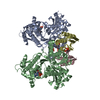
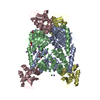
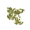
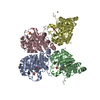
-
Structure visualization

Structure viewerMolecule:
MolmilJmol/JSmol

Downloads & links

-
Assembly

Deposited unit
A: MAJOR DNA-BINDING PROTEIN
B: MAJOR DNA-BINDING PROTEIN
hetero molecules


Theoretical massNumber of molelcules
Total (without water)246,27112
Polymers244,5362
Non-polymers1,73610
Water1,71195
1
A: MAJOR DNA-BINDING PROTEIN
hetero molecules


Theoretical massNumber of molelcules
Total (without water)123,1366
Polymers122,2681
Non-polymers8685
Water181
TypeNameSymmetry operationNumber
identity operation1_555x,y,z1
MethodPQS
2
B: MAJOR DNA-BINDING PROTEIN
hetero molecules


Theoretical massNumber of molelcules
Total (without water)123,1366
Polymers122,2681
Non-polymers8685
Water181
TypeNameSymmetry operationNumber
identity operation1_555x,y,z1
MethodPQS
Unit cell
Length a, b, c (Å)100.910, 145.370, 162.030
Angle α, β, γ (deg.)90.00, 90.00, 90.00
Int Tables number19
Space group name H-MP212121

-
Components

#1: Protein MAJOR DNA-BINDING PROTEIN / SINGLE-STRANDED DNA BINDING PROTEIN / INFECTED CELL PROTEIN 8 / ICP 8 PROTEIN


Mass: 122267.797 Da / Num. of mol.: 2 / Mutation: YES
Source method: isolated from a genetically manipulated source
Source: (gene. exp.) HUMAN HERPESVIRUS 1 (Herpes simplex virus type 1)
Plasmid: PE29 / Cell line (production host): High Five / Production host: Trichoplusia ni (cabbage looper) / References: UniProt: P04296
#2: Chemical ChemComp-ZN / ZINC ION


Mass: 65.409 Da / Num. of mol.: 2 / Source method: obtained synthetically / Formula: Zn
#3: Chemical
ChemComp-HG / MERCURY (II) ION / Mercury (element)


Mass: 200.590 Da / Num. of mol.: 8 / Source method: obtained synthetically / Formula: Hg
#4: Water ChemComp-HOH / water / Water


Mass: 18.015 Da / Num. of mol.: 95 / Source method: isolated from a natural source / Formula: H2O
Compound detailsSINGLE-STRAND DNA-BINDING PROTEIN REQUIRED FOR DNA REPLICATION. AS VIRAL DNA REPLICATION PROCEEDS, ...SINGLE-STRAND DNA-BINDING PROTEIN REQUIRED FOR DNA REPLICATION. AS VIRAL DNA REPLICATION PROCEEDS, IT MIGRATES TO GLOBULAR INTRANUCLEAR STRUCTURES (REPLICATION COMPARTMENTS). ENGINEERED MUTATION IN CHAIN A, CYS 254 TO SER 254 ENGINEERED MUTATION IN CHAIN B, CYS 254 TO SER 254 ENGINEERED MUTATION IN CHAIN A, CYS 455 TO SER 455 ENGINEERED MUTATION IN CHAIN B, CYS 455 TO SER 455

-
Experimental details

-
Experiment

ExperimentMethod: X-RAY DIFFRACTION / Number of used crystals: 1

-
Sample preparation

CrystalDensity Matthews: 2.4 Å3/Da / Density % sol: 49 %
Crystal growpH: 6
Details: 12% PEG 3K, 0.1 M SODIUM/POTASSIUM PHASPHATE PH 6.0

-
Data collection

DiffractionMean temperature: 100 K
Diffraction sourceSource: SYNCHROTRON / Site: EMBL/DESY, HAMBURG / Beamline: BW7B / Wavelength: 0.8467
DetectorType: MARRESEARCH / Detector: CCD / Date: Mar 15, 1999 / Details: MIRRORS
RadiationMonochromator: SILICON / Protocol: SINGLE WAVELENGTH / Monochromatic (M) / Laue (L): M / Scattering type: x-ray
Radiation wavelengthWavelength: 0.8467 Å / Relative weight: 1
ReflectionResolution: 3→30 Å / Num. obs: 47265 / % possible obs: 98 % / Redundancy: 3.9 % / Rmerge(I) obs: 0.09 / Net I/σ(I): 9.8
Reflection shellResolution: 3→20 Å / Redundancy: 3.6 % / Rmerge(I) obs: 0.3 / Mean I/σ(I) obs: 2.98 / % possible all: 97

-
Processing

Software
NameVersionClassification
DENZOdata reduction
SCALEPACKdata scaling
MLPHAREphasing
SHELXDphasing
CNS1.1refinement
RefinementMethod to determine structure: MAD / Resolution: 3→20 Å / Cross valid method: THROUGHOUT / σ(F): 0
RfactorNum. reflection% reflectionSelection details
Rfree0.2857 1187 2.5 %RANDOM
Rwork0.235 ---
obs0.235 47263 98 %-
Displacement parametersBiso mean: 35.8 Å2
Baniso -1Baniso -2Baniso -3
1--15.075 Å20 Å20 Å2
2--10.603 Å20 Å2
3---4.472 Å2
Refinement stepCycle: LAST / Resolution: 3→20 Å
ProteinNucleic acidLigandSolventTotal
Num. atoms15693 0 10 95 15798
Refine LS restraints
Refine-IDTypeDev ideal
X-RAY DIFFRACTIONc_bond_d0.0079
X-RAY DIFFRACTIONc_bond_d_na
X-RAY DIFFRACTIONc_bond_d_prot
X-RAY DIFFRACTIONc_angle_d
X-RAY DIFFRACTIONc_angle_d_na
X-RAY DIFFRACTIONc_angle_d_prot
X-RAY DIFFRACTIONc_angle_deg1.36
X-RAY DIFFRACTIONc_angle_deg_na
X-RAY DIFFRACTIONc_angle_deg_prot
X-RAY DIFFRACTIONc_dihedral_angle_d
X-RAY DIFFRACTIONc_dihedral_angle_d_na
X-RAY DIFFRACTIONc_dihedral_angle_d_prot
X-RAY DIFFRACTIONc_improper_angle_d
X-RAY DIFFRACTIONc_improper_angle_d_na
X-RAY DIFFRACTIONc_improper_angle_d_prot
X-RAY DIFFRACTIONc_mcbond_it
X-RAY DIFFRACTIONc_mcangle_it
X-RAY DIFFRACTIONc_scbond_it
X-RAY DIFFRACTIONc_scangle_it
Refine LS restraints NCSNCS model details: RESTRAINTS / Rms dev Biso : 13.24 Å2 / Rms dev position: 2 Å / Weight Biso : 1 / Weight position: 300
LS refinement shellResolution: 3→20 Å / Total num. of bins used: 23 /
Num. reflection% reflection
Rwork1920 -
obs-97 %

+
About Yorodumi

-
News

-
Feb 9, 2022. New format data for meta-information of EMDB entries

New format data for meta-information of EMDB entries

  • Version 3 of the EMDB header file is now the official format.
  • The previous official version 1.9 will be removed from the archive.

Related info.:EMDB header

External links:wwPDB to switch to version 3 of the EMDB data model

-
Aug 12, 2020. Covid-19 info

Covid-19 info

URL: https://pdbj.org/emnavi/covid19.php

New page: Covid-19 featured information page in EM Navigator.

Related info.:Covid-19 info / Mar 5, 2020. Novel coronavirus structure data

+
Mar 5, 2020. Novel coronavirus structure data

Novel coronavirus structure data

Related info.:Yorodumi Speices / Aug 12, 2020. Covid-19 info

External links:COVID-19 featured content - PDBj / Molecule of the Month (242):Coronavirus Proteases

+
Jan 31, 2019. EMDB accession codes are about to change! (news from PDBe EMDB page)

EMDB accession codes are about to change! (news from PDBe EMDB page)

  • The allocation of 4 digits for EMDB accession codes will soon come to an end. Whilst these codes will remain in use, new EMDB accession codes will include an additional digit and will expand incrementally as the available range of codes is exhausted. The current 4-digit format prefixed with “EMD-” (i.e. EMD-XXXX) will advance to a 5-digit format (i.e. EMD-XXXXX), and so on. It is currently estimated that the 4-digit codes will be depleted around Spring 2019, at which point the 5-digit format will come into force.
  • The EM Navigator/Yorodumi systems omit the EMD- prefix.

Related info.:Q: What is EMD? / ID/Accession-code notation in Yorodumi/EM Navigator

External links:EMDB Accession Codes are Changing Soon! / Contact to PDBj

+
Jul 12, 2017. Major update of PDB

Major update of PDB

  • wwPDB released updated PDB data conforming to the new PDBx/mmCIF dictionary.
  • This is a major update changing the version number from 4 to 5, and with Remediation, in which all the entries are updated.
  • In this update, many items about electron microscopy experimental information are reorganized (e.g. em_software).
  • Now, EM Navigator and Yorodumi are based on the updated data.

External links:wwPDB Remediation / Enriched Model Files Conforming to OneDep Data Standards Now Available in the PDB FTP Archive

-
Yorodumi

Thousand views of thousand structures

  • Yorodumi is a browser for structure data from EMDB, PDB, SASBDB, etc.
  • This page is also the successor to EM Navigator detail page, and also detail information page/front-end page for Omokage search.
  • The word "yorodu" (or yorozu) is an old Japanese word meaning "ten thousand". "mi" (miru) is to see.

Related info.:EMDB / PDB / SASBDB / Comparison of 3 databanks / Yorodumi Search / Aug 31, 2016. New EM Navigator & Yorodumi / Yorodumi Papers / Jmol/JSmol / Function and homology information / Changes in new EM Navigator and Yorodumi

Read more