[English] 日本語
Yorodumi
- PDB-2wp0: Crystal structure of a HobA-DnaA (domain I-II) complex from Helic... -

+
Open data


ID or keywords:

Loading...

-
Basic information

Entry
Database: PDB / ID: 2wp0
Title
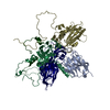
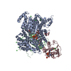
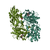
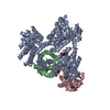
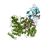
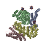
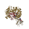
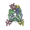
Crystal structure of a HobA-DnaA (domain I-II) complex from Helicobacter pylori.
Components
  • CHROMOSOMAL REPLICATION INITIATOR PROTEIN DNAA
  • HOBA
KeywordsDNA BINDING PROTEIN / NUCLEOTIDE-BINDING / DNA REPLICATION INITIATION / DIAA / HOBA / DNAA / ATP-BINDING
Function / homology
Function and homology information


DNA replication origin binding / regulation of DNA replication / DNA replication initiation / DNA replication / ATP binding / identical protein binding / metal ion binding / plasma membrane / cytoplasm
Similarity search - Function
DNA replication regulator HobA / DNA replication regulator, HobA / DNA replication regulator HobA superfamily / DNA replication regulator / DnaA, N-terminal domain / DnaA, N-terminal domain superfamily / Chromosomal replication control, initiator DnaA / Chromosomal replication initiator, DnaA C-terminal / Chromosomal replication control, initiator DnaA, conserved site / Bacterial dnaA protein helix-turn-helix ...DNA replication regulator HobA / DNA replication regulator, HobA / DNA replication regulator HobA superfamily / DNA replication regulator / DnaA, N-terminal domain / DnaA, N-terminal domain superfamily / Chromosomal replication control, initiator DnaA / Chromosomal replication initiator, DnaA C-terminal / Chromosomal replication control, initiator DnaA, conserved site / Bacterial dnaA protein helix-turn-helix / DnaA protein signature. / Bacterial dnaA protein helix-turn-helix domain / Chromosomal replication control, initiator DnaA-like / Chromosomal replication initiator protein DnaA / Bacterial dnaA protein / Trp repressor/replication initiator / GMP Synthetase; Chain A, domain 3 / ATPases associated with a variety of cellular activities / AAA+ ATPase domain / P-loop containing nucleoside triphosphate hydrolase / Rossmann fold / 2-Layer Sandwich / 3-Layer(aba) Sandwich / Alpha Beta
Similarity search - Domain/homology
ACETATE ION / DNA replication regulator family protein / Chromosomal replication initiator protein DnaA
Similarity search - Component
Biological speciesHELICOBACTER PYLORI (bacteria)
MethodX-RAY DIFFRACTION / SYNCHROTRON / MOLECULAR REPLACEMENT / Resolution: 2.67 Å
AuthorsNatrajan, G. / Noirot-Gros, M.-F. / Zawilak-Pawlik, A. / Kapp, U. / Terradot, L.
CitationJournal: Proc.Natl.Acad.Sci.USA / Year: 2009
Title: The Structure of a Dnaa/Hoba Complex from Helicobacter Pylori Provides Insight Into Regulation of DNA Replication in Bacteria.
Authors: Natrajan, G. / Noirot-Gros, M.F. / Zawilak-Pawlik, A. / Kapp, U. / Terradot, L.
History
DepositionJul 31, 2009Deposition site: PDBE / Processing site: PDBE
Revision 1.0Nov 17, 2009Provider: repository / Type: Initial release
Revision 1.1May 8, 2011Group: Version format compliance
Revision 1.2Jul 13, 2011Group: Version format compliance
Revision 1.3Jul 12, 2017Group: Derived calculations / Category: pdbx_struct_conn_angle / struct_conn
Revision 1.4Dec 20, 2023Group: Data collection / Database references ...Data collection / Database references / Derived calculations / Other / Refinement description
Category: chem_comp_atom / chem_comp_bond ...chem_comp_atom / chem_comp_bond / database_2 / pdbx_database_status / pdbx_initial_refinement_model / struct_conn
Item: _database_2.pdbx_DOI / _database_2.pdbx_database_accession ..._database_2.pdbx_DOI / _database_2.pdbx_database_accession / _pdbx_database_status.status_code_sf / _struct_conn.pdbx_dist_value / _struct_conn.ptnr1_auth_asym_id / _struct_conn.ptnr1_auth_comp_id / _struct_conn.ptnr1_auth_seq_id / _struct_conn.ptnr1_label_asym_id / _struct_conn.ptnr1_label_atom_id / _struct_conn.ptnr1_label_comp_id / _struct_conn.ptnr1_label_seq_id / _struct_conn.ptnr1_symmetry / _struct_conn.ptnr2_auth_asym_id / _struct_conn.ptnr2_auth_comp_id / _struct_conn.ptnr2_auth_seq_id / _struct_conn.ptnr2_label_asym_id / _struct_conn.ptnr2_label_atom_id / _struct_conn.ptnr2_label_comp_id / _struct_conn.ptnr2_label_seq_id / _struct_conn.ptnr2_symmetry

-
Structure visualization

Structure viewerMolecule:
MolmilJmol/JSmol

Downloads & links

-
Assembly

Deposited unit
A: HOBA
B: HOBA
C: CHROMOSOMAL REPLICATION INITIATOR PROTEIN DNAA
D: CHROMOSOMAL REPLICATION INITIATOR PROTEIN DNAA
hetero molecules


Theoretical massNumber of molelcules
Total (without water)68,05224
Polymers67,3434
Non-polymers70920
Water1,928107
1
A: HOBA
B: HOBA
C: CHROMOSOMAL REPLICATION INITIATOR PROTEIN DNAA
D: CHROMOSOMAL REPLICATION INITIATOR PROTEIN DNAA
hetero molecules

A: HOBA
B: HOBA
C: CHROMOSOMAL REPLICATION INITIATOR PROTEIN DNAA
D: CHROMOSOMAL REPLICATION INITIATOR PROTEIN DNAA
hetero molecules


Theoretical massNumber of molelcules
Total (without water)136,10448
Polymers134,6878
Non-polymers1,41840
Water1448
TypeNameSymmetry operationNumber
identity operation1_555x,y,z1
crystal symmetry operation2_555-x,y,-z1
Buried area22070 Å2
ΔGint-381.17 kcal/mol
Surface area48030 Å2
MethodPISA
Unit cell
Length a, b, c (Å)126.560, 55.500, 96.830
Angle α, β, γ (deg.)90.00, 97.55, 90.00
Int Tables number5
Space group name H-MC121
Noncrystallographic symmetry (NCS)NCS domain:
IDEns-ID
11
21
12
22

NCS domain segments:
Dom-IDComponent-IDEns-IDSelection details
111CHAIN A AND (RESSEQ 1:180 )
211CHAIN B AND (RESSEQ 1:180 )
112CHAIN C AND (RESSEQ 10:74 OR RESSEQ 80:91 )
212CHAIN D AND (RESSEQ 10:74 OR RESSEQ 80:91 )

NCS ensembles :
ID
1
2

-
Components

-
Protein , 2 types, 4 molecules ABCD

#1: Protein HOBA


Mass: 21039.197 Da / Num. of mol.: 2
Source method: isolated from a genetically manipulated source
Details: HOBA PROTEIN (HP 1230) / Source: (gene. exp.) HELICOBACTER PYLORI (bacteria) / Production host: ESCHERICHIA COLI (E. coli) / Strain (production host): BL21(DE3) / References: UniProt: O25828
#2: Protein CHROMOSOMAL REPLICATION INITIATOR PROTEIN DNAA


Mass: 12632.481 Da / Num. of mol.: 2 / Fragment: DOMAIN I-II, RESIDUES 1-120
Source method: isolated from a genetically manipulated source
Details: N-TERMINAL FRAGMENT OF DNAA / Source: (gene. exp.) HELICOBACTER PYLORI (bacteria) / Production host: ESCHERICHIA COLI (E. coli) / Strain (production host): BL21(DE3) / References: UniProt: O26057

-
Non-polymers , 5 types, 127 molecules

#3: Chemical
ChemComp-CL / CHLORIDE ION / Chloride


Mass: 35.453 Da / Num. of mol.: 6 / Source method: obtained synthetically / Formula: Cl
#4: Chemical ChemComp-GOL / GLYCEROL / GLYCERIN / PROPANE-1,2,3-TRIOL / Glycerol


Mass: 92.094 Da / Num. of mol.: 2 / Source method: obtained synthetically / Formula: C3H8O3
#5: Chemical
ChemComp-NA / SODIUM ION


Mass: 22.990 Da / Num. of mol.: 11 / Source method: obtained synthetically / Formula: Na
#6: Chemical ChemComp-ACT / ACETATE ION / Acetate


Mass: 59.044 Da / Num. of mol.: 1 / Source method: obtained synthetically / Formula: C2H3O2
#7: Water ChemComp-HOH / water / Water


Mass: 18.015 Da / Num. of mol.: 107 / Source method: isolated from a natural source / Formula: H2O

-
Details

Sequence detailsTHE ACCESSION NUMBER IS FOR THE FULL LENGTH PROTEIN

-
Experimental details

-
Experiment

ExperimentMethod: X-RAY DIFFRACTION / Number of used crystals: 1

-
Sample preparation

CrystalDensity Matthews: 2.63 Å3/Da / Density % sol: 53.3 %
Description: A COMBINATION OF 2UVP FOR HOBA AND A HOMOLGY MODEL FOR DNAA I-II WAS USED FOR MOLECULAR REPLACEMENT.
Crystal growpH: 8
Details: 100 MM TRIS-HCL PH 8.0, 200 MM K-ACETATE, 19-22% PEG 3350

-
Data collection

DiffractionMean temperature: 100 K
Diffraction sourceSource: SYNCHROTRON / Site: ESRF / Beamline: ID14-4 / Wavelength: 0.91
DetectorType: ADSC CCD / Detector: CCD / Date: Aug 17, 2008 / Details: MIRRORS
RadiationMonochromator: SI 111 / Protocol: SINGLE WAVELENGTH / Monochromatic (M) / Laue (L): M / Scattering type: x-ray
Radiation wavelengthWavelength: 0.91 Å / Relative weight: 1
ReflectionResolution: 2.67→50.77 Å / Num. obs: 17498 / % possible obs: 93.4 % / Observed criterion σ(I): 1.1 / Redundancy: 2.6 % / Biso Wilson estimate: 39.3 Å2 / Rmerge(I) obs: 0.12 / Net I/σ(I): 6.8
Reflection shellResolution: 2.67→2.81 Å / Redundancy: 2.5 % / Rmerge(I) obs: 0.55 / Mean I/σ(I) obs: 1.7 / % possible all: 94.7

-
Processing

Software
NameVersionClassification
PHENIX(PHENIX.REFINE)refinement
iMOSFLMdata reduction
SCALAdata scaling
PHASERphasing
RefinementMethod to determine structure: MOLECULAR REPLACEMENT
Starting model: PDB ENTRY 2UVP
Resolution: 2.67→50.756 Å / SU ML: 0.41 / σ(F): 1.34 / Phase error: 27.95 / Stereochemistry target values: ML / Details: RESIDUE 75 MISSING FROM CHAIN C.
RfactorNum. reflection% reflection
Rfree0.2663 897 5.17 %
Rwork0.2262 --
obs0.2283 17357 90.41 %
Solvent computationShrinkage radii: 0.9 Å / VDW probe radii: 1.11 Å / Solvent model: FLAT BULK SOLVENT MODEL / Bsol: 60 Å2 / ksol: 0.375 e/Å3
Displacement parameters
Baniso -1Baniso -2Baniso -3
1--4.9313 Å2-0 Å20.9037 Å2
2---1.8336 Å2-0 Å2
3---9.8006 Å2
Refinement stepCycle: LAST / Resolution: 2.67→50.756 Å
ProteinNucleic acidLigandSolventTotal
Num. atoms4339 0 33 107 4479
Refine LS restraints
Refine-IDTypeDev idealNumber
X-RAY DIFFRACTIONf_bond_d0.0114451
X-RAY DIFFRACTIONf_angle_d1.5066019
X-RAY DIFFRACTIONf_dihedral_angle_d21.2441610
X-RAY DIFFRACTIONf_chiral_restr0.117675
X-RAY DIFFRACTIONf_plane_restr0.006751
Refine LS restraints NCS
Ens-IDDom-IDAuth asym-IDNumberRefine-IDTypeRms dev position (Å)
11A1487X-RAY DIFFRACTIONPOSITIONAL
12B1487X-RAY DIFFRACTIONPOSITIONAL0.273
21C611X-RAY DIFFRACTIONPOSITIONAL
22D611X-RAY DIFFRACTIONPOSITIONAL0.691
LS refinement shell
Resolution (Å)Rfactor RfreeNum. reflection RfreeRfactor RworkNum. reflection RworkRefine-ID% reflection obs (%)
2.67-2.83730.38431500.31352667X-RAY DIFFRACTION90
2.8373-3.05630.33771550.29182820X-RAY DIFFRACTION94
3.0563-3.36380.30951700.24722816X-RAY DIFFRACTION94
3.3638-3.85040.23191460.21692813X-RAY DIFFRACTION92
3.8504-4.85060.21341430.17342739X-RAY DIFFRACTION90
4.8506-50.76480.19921330.18392605X-RAY DIFFRACTION83
Refinement TLS params.

Method: refined / Refine-ID: X-RAY DIFFRACTION

IDL112)L122)L132)L222)L232)L332)S11 (Å °)S12 (Å °)S13 (Å °)S21 (Å °)S22 (Å °)S23 (Å °)S31 (Å °)S32 (Å °)S33 (Å °)T112)T122)T132)T222)T232)T332)Origin x (Å)Origin y (Å)Origin z (Å)
10.7171-0.0739-0.43820.2752-0.15940.94910.06470.02150.11140.06910.0456-0.0217-0.06190.07-0.11370.1875-0.0183-0.01960.1473-0.02710.236217.0163-17.204310.0061
20.5651-0.05960.53180.3110.0330.95860.06570.0093-0.0381-0.07370.0487-0.05680.05250.0627-0.11050.19570.00560.01180.141-0.03360.241916.8997-26.2902-9.9762
30.39520.54210.25710.77530.0721.66720.0981-0.0228-0.4194-0.0391-0.3596-0.58490.1988-0.19150.0710.252-0.102-0.11650.27060.11270.379412.8329-43.999232.8747
40.4787-0.2443-0.24170.1633-0.05771.03880.052-0.07830.2424-0.089-0.0757-0.2727-0.315-0.1912-0.03990.24660.06580.07380.17990.04110.248512.64150.8027-32.8355
Refinement TLS group
IDRefine-IDRefine TLS-IDSelection details
1X-RAY DIFFRACTION1CHAIN A
2X-RAY DIFFRACTION2CHAIN B
3X-RAY DIFFRACTION3CHAIN C
4X-RAY DIFFRACTION4CHAIN D

+
About Yorodumi

-
News

-
Feb 9, 2022. New format data for meta-information of EMDB entries

New format data for meta-information of EMDB entries

  • Version 3 of the EMDB header file is now the official format.
  • The previous official version 1.9 will be removed from the archive.

Related info.:EMDB header

External links:wwPDB to switch to version 3 of the EMDB data model

-
Aug 12, 2020. Covid-19 info

Covid-19 info

URL: https://pdbj.org/emnavi/covid19.php

New page: Covid-19 featured information page in EM Navigator.

Related info.:Covid-19 info / Mar 5, 2020. Novel coronavirus structure data

+
Mar 5, 2020. Novel coronavirus structure data

Novel coronavirus structure data

Related info.:Yorodumi Speices / Aug 12, 2020. Covid-19 info

External links:COVID-19 featured content - PDBj / Molecule of the Month (242):Coronavirus Proteases

+
Jan 31, 2019. EMDB accession codes are about to change! (news from PDBe EMDB page)

EMDB accession codes are about to change! (news from PDBe EMDB page)

  • The allocation of 4 digits for EMDB accession codes will soon come to an end. Whilst these codes will remain in use, new EMDB accession codes will include an additional digit and will expand incrementally as the available range of codes is exhausted. The current 4-digit format prefixed with “EMD-” (i.e. EMD-XXXX) will advance to a 5-digit format (i.e. EMD-XXXXX), and so on. It is currently estimated that the 4-digit codes will be depleted around Spring 2019, at which point the 5-digit format will come into force.
  • The EM Navigator/Yorodumi systems omit the EMD- prefix.

Related info.:Q: What is EMD? / ID/Accession-code notation in Yorodumi/EM Navigator

External links:EMDB Accession Codes are Changing Soon! / Contact to PDBj

+
Jul 12, 2017. Major update of PDB

Major update of PDB

  • wwPDB released updated PDB data conforming to the new PDBx/mmCIF dictionary.
  • This is a major update changing the version number from 4 to 5, and with Remediation, in which all the entries are updated.
  • In this update, many items about electron microscopy experimental information are reorganized (e.g. em_software).
  • Now, EM Navigator and Yorodumi are based on the updated data.

External links:wwPDB Remediation / Enriched Model Files Conforming to OneDep Data Standards Now Available in the PDB FTP Archive

-
Yorodumi

Thousand views of thousand structures

  • Yorodumi is a browser for structure data from EMDB, PDB, SASBDB, etc.
  • This page is also the successor to EM Navigator detail page, and also detail information page/front-end page for Omokage search.
  • The word "yorodu" (or yorozu) is an old Japanese word meaning "ten thousand". "mi" (miru) is to see.

Related info.:EMDB / PDB / SASBDB / Comparison of 3 databanks / Yorodumi Search / Aug 31, 2016. New EM Navigator & Yorodumi / Yorodumi Papers / Jmol/JSmol / Function and homology information / Changes in new EM Navigator and Yorodumi

Read more