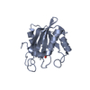
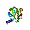
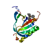
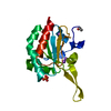
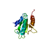
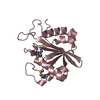
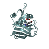
Journal: Sci Rep / Year: 2014 Title: Structural insight into substrate recognition by the endoplasmic reticulum folding-sensor enzyme: crystal structure of third thioredoxin-like domain of UDP-glucose:glycoprotein glucosyltransferase Authors: Zhu, T. / Satoh, T. / Kato, K.
Monochromator: Si111 / Protocol: SINGLE WAVELENGTH / Monochromatic (M) / Laue (L): M / Scattering type: x-ray
Radiation wavelength
Wavelength: 0.97921 Å / Relative weight: 1
Reflection
Resolution: 1.7→50 Å / Num. all: 20126 / Num. obs: 19829 / % possible obs: 98.5 % / Observed criterion σ(I): -3 / Redundancy: 6.8 % / Biso Wilson estimate: 17.6 Å2 / Rmerge(I) obs: 0.082 / Net I/σ(I): 47.9
Reflection shell
Resolution: 1.7→1.73 Å / Redundancy: 6.8 % / Rmerge(I) obs: 0.366 / Mean I/σ(I) obs: 7.2 / Num. unique all: 970 / % possible all: 98.9
-
Processing
Software
Name
Version
Classification
HKL-2000
datacollection
SHELXS
phasing
REFMAC
5.8.0069
refinement
DENZO
datareduction
SCALEPACK
datascaling
Refinement
Method to determine structure: SAD / Resolution: 1.7→20 Å / Cor.coef. Fo:Fc: 0.952 / Cor.coef. Fo:Fc free: 0.926 / SU B: 2.065 / SU ML: 0.07 / Cross valid method: THROUGHOUT / ESU R: 0.112 / ESU R Free: 0.115 / Stereochemistry target values: MAXIMUM LIKELIHOOD / Details: HYDROGENS HAVE BEEN ADDED IN THE RIDING POSITIONS
Rfactor
Num. reflection
% reflection
Selection details
Rfree
0.247
976
5 %
RANDOM
Rwork
0.203
-
-
-
obs
0.205
18450
97.39 %
-
all
-
18450
-
-
Solvent computation
Ion probe radii: 0.8 Å / Shrinkage radii: 0.8 Å / VDW probe radii: 1.2 Å / Solvent model: MASK
 Movie
Movie Controller


 Open data
 Basic information
 Components
 Keywords
 Function and homology information
 Chaetomium thermophilum var. thermophilum DSM 1495 (fungus)
 X-RAY DIFFRACTION /
  Authors
 Citation
 Structure visualization
 Downloads & links
 Other downloads
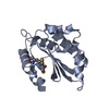


 PDBj
PDBj


 Assembly

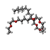
 Mass: 536.782 Da / Num. of mol.: 1 / Source method: obtained synthetically / Formula: C29H60O8
 Mass: 18.015 Da / Num. of mol.: 120 / Source method: isolated from a natural source / Formula: H2O
 Sample preparation
 / Beamline: AR-NW12A / Wavelength: 0.97921 Å
 Processing