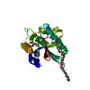
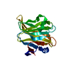
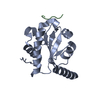
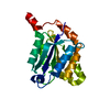
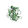
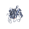
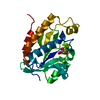
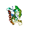
- PDB-4m90: crystal structure of oxidized hN33/Tusc3 -
+
Open data
ID or keywords:
Loading...
-
Basic information
Entry
Database: PDB / ID: 4m90
Title
crystal structure of oxidized hN33/Tusc3
Components
Tumor suppressor candidate 3
Keywords
OXIDOREDUCTASE / Thioredoxin-like fold / Redox active / endoplasmic reticulum
Function / homology
Function and homology information
Asparagine N-linked glycosylation / magnesium ion transport / Miscellaneous transport and binding events / magnesium ion transmembrane transport / oligosaccharyltransferase complex / : / magnesium ion transmembrane transporter activity / protein N-linked glycosylation / transmembrane transport / cognition ...Asparagine N-linked glycosylation / magnesium ion transport / Miscellaneous transport and binding events / magnesium ion transmembrane transport / oligosaccharyltransferase complex / : / magnesium ion transmembrane transporter activity / protein N-linked glycosylation / transmembrane transport / cognition / Maturation of spike protein / endoplasmic reticulum membrane / mitochondrion / plasma membrane Similarity search - Function
Resolution: 1.6→32.32 Å / Cor.coef. Fo:Fc: 0.959 / Cor.coef. Fo:Fc free: 0.944 / SU B: 3.942 / SU ML: 0.066 / Cross valid method: THROUGHOUT / ESU R: 0.092 / ESU R Free: 0.093 / Stereochemistry target values: MAXIMUM LIKELIHOOD / Details: HYDROGENS HAVE BEEN ADDED IN THE RIDING POSITIONS
Rfactor
Num. reflection
% reflection
Selection details
Rfree
0.22003
443
2.1 %
RANDOM
Rwork
0.18484
-
-
-
obs
0.18552
20596
98.88 %
-
all
-
20596
-
-
Solvent computation
Ion probe radii: 0.8 Å / Shrinkage radii: 0.8 Å / VDW probe radii: 1.2 Å / Solvent model: MASK
 Movie
Movie Controller


 Open data
 Basic information
 Components
 Keywords
 Function and homology information
 Homo sapiens (human)
 X-RAY DIFFRACTION /
  Authors
 Citation
 Structure visualization
 Downloads & links
 Other downloads


 PDBj
PDBj
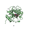
 Assembly
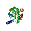

 Mass: 18.015 Da / Num. of mol.: 136 / Source method: isolated from a natural source / Formula: H2O
 Sample preparation
 / Beamline: X06SA / Wavelength: 1 Å
 Processing