 Movie
Movie Controller

[English] 日本語
 Yorodumi
- PDB-5zhg: Human group C rotavirus VP8*s recognize type A histo-blood group ... -
+ Open data

- Basic information
| Entry | Database: PDB / ID: 5zhg | ||||||||||||||||||||||||||||
|---|---|---|---|---|---|---|---|---|---|---|---|---|---|---|---|---|---|---|---|---|---|---|---|---|---|---|---|---|---|
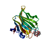
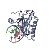
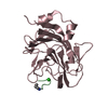
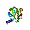
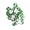
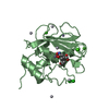
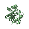
| Title | Human group C rotavirus VP8*s recognize type A histo-blood group antigens as ligands | ||||||||||||||||||||||||||||
 Components Components | Outer capsid protein VP8* Keywords KeywordsVIRAL PROTEIN / group C rotavirus / VP8* / galactin-like / histo-blood group antigen. | Function / homology |  Function and homology information Function and homology informationhost cell rough endoplasmic reticulum / permeabilization of host organelle membrane involved in viral entry into host cell / viral outer capsid / host cell endoplasmic reticulum-Golgi intermediate compartment / virion attachment to host cell / host cell plasma membrane Similarity search - Function Biological species |  Human rotavirus C Human rotavirus CMethod |  X-RAY DIFFRACTION / Resolution: 1.799 Å X-RAY DIFFRACTION / Resolution: 1.799 Å  Authors AuthorsSun, X. / Duan, Z. | Funding support | |  China, 1items China, 1items
 CitationJournal: J. Virol. / Year: 2018 CitationJournal: J. Virol. / Year: 2018Title: Human Group C Rotavirus VP8*s Recognize Type A Histo-Blood Group Antigens as Ligands. Authors: Sun, X. / Wang, L. / Qi, J. / Li, D. / Wang, M. / Cong, X. / Peng, R. / Chai, W. / Zhang, Q. / Wang, H. / Wen, H. / Gao, G.F. / Tan, M. / Duan, Z. History |
|
- Structure visualization
| Structure viewer | Molecule: MolmilJmol/JSmol |
|---|
- Downloads & links
-Download
| PDBx/mmCIF format | 5zhg.cif.gz | 87.6 KB | Display | PDBx/mmCIF format |
|---|---|---|---|---|
| PDB format | pdb5zhg.ent.gz | 65.9 KB | Display | PDB format |
| PDBx/mmJSON format | 5zhg.json.gz | Tree view | PDBx/mmJSON format | |
| Others |  Other downloads Other downloads |
-Validation report
| Arichive directory | https://data.pdbj.org/pub/pdb/validation_reports/zh/5zhgftp://data.pdbj.org/pub/pdb/validation_reports/zh/5zhg | HTTPS FTP |
|---|
-Related structure data


-Links
 PDBj
PDBj- Assembly
| Deposited unit | 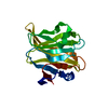
| ||||||||
|---|---|---|---|---|---|---|---|---|---|
| 1 |
| ||||||||
| Unit cell |
|
-Components
| #1: Protein | Mass: 18738.826 Da / Num. of mol.: 1 Source method: isolated from a genetically manipulated source Source: (gene. exp.) Human rotavirus CProduction host: References: UniProt: Q82040 |
|---|---|
| #2: Water | ChemComp-HOH /  Mass: 18.015 Da / Num. of mol.: 208 / Source method: isolated from a natural source / Formula: H2O Mass: 18.015 Da / Num. of mol.: 208 / Source method: isolated from a natural source / Formula: H2O |
-Experimental details
-Experiment
| Experiment | Method: X-RAY DIFFRACTION / Number of used crystals: 1 |
|---|
- Sample preparation
| Crystal | Density Matthews: 2.52 Å3/Da / Density % sol: 51.26 % |
|---|---|
| Crystal grow | Temperature: 291 K / Method: evaporation / pH: 6.5 Details: 0.1 M Sodium chloride, 0.1 M BIS-TRIS pH 6.5, 1.5 M Ammonium sulfate |
-Data collection
| Diffraction | Mean temperature: 113 K |
|---|---|
| Diffraction source | Source: SEALED TUBE / Type: Xenocs GeniX 3D Cu HF / Wavelength: 1.54178 Å |
| Detector | Type: RIGAKU RAXIS IV / Detector: IMAGE PLATE / Date: Aug 31, 2017 |
| Radiation | Protocol: SINGLE WAVELENGTH / Monochromatic (M) / Laue (L): M / Scattering type: x-ray |
| Radiation wavelength | Wavelength: 1.54178 Å / Relative weight: 1 |
| Reflection | Resolution: 1.799→50 Å / Num. obs: 18290 / % possible obs: 99.4 % / Redundancy: 10.3 % / Biso Wilson estimate: 20.43 Å2 / Rmerge(I) obs: 0.099 / Net I/σ(I): 21.019 |
| Reflection shell | Resolution: 1.8→1.86 Å / Rmerge(I) obs: 0.565 |
- Processing
| Software |
| ||||||||||||||||||||||||||||||||||||||||||||||||||||||||
|---|---|---|---|---|---|---|---|---|---|---|---|---|---|---|---|---|---|---|---|---|---|---|---|---|---|---|---|---|---|---|---|---|---|---|---|---|---|---|---|---|---|---|---|---|---|---|---|---|---|---|---|---|---|---|---|---|---|
| Refinement | Resolution: 1.799→38.129 Å / SU ML: 0.22 / Cross valid method: THROUGHOUT / σ(F): 1.35 / Phase error: 20.93
| ||||||||||||||||||||||||||||||||||||||||||||||||||||||||
| Solvent computation | Shrinkage radii: 0.9 Å / VDW probe radii: 1.11 Å | ||||||||||||||||||||||||||||||||||||||||||||||||||||||||
| Displacement parameters | Biso max: 89.49 Å2 / Biso mean: 23.6151 Å2 / Biso min: 10.07 Å2 | ||||||||||||||||||||||||||||||||||||||||||||||||||||||||
| Refinement step | Cycle: final / Resolution: 1.799→38.129 Å
| ||||||||||||||||||||||||||||||||||||||||||||||||||||||||
| Refine LS restraints |
| ||||||||||||||||||||||||||||||||||||||||||||||||||||||||
| LS refinement shell | Refine-ID: X-RAY DIFFRACTION / Rfactor Rfree error: 0 / Total num. of bins used: 7
| ||||||||||||||||||||||||||||||||||||||||||||||||||||||||
| Refinement TLS params. | Method: refined / Origin x: 39.6356 Å / Origin y: 55.8409 Å / Origin z: 139.6974 Å
| ||||||||||||||||||||||||||||||||||||||||||||||||||||||||
| Refinement TLS group | Selection details: all |
