 Movie
Movie Controller

[English] 日本語
 Yorodumi
- PDB-4djj: Crystal structure of the complex of Peptidyl-tRNA hydrolase from ... -
+ Open data

- Basic information
| Entry | Database: PDB / ID: 4djj | ||||||
|---|---|---|---|---|---|---|---|
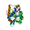
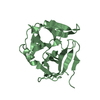
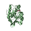
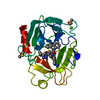
| Title | Crystal structure of the complex of Peptidyl-tRNA hydrolase from Pseudomonas aeruginosa with Pimelic acid at 2.9 Angstrom resolution | ||||||
 Components Components | Peptidyl-tRNA hydrolase | ||||||
 Keywords Keywords | HYDROLASE / esterase | ||||||
| Function / homology |  Function and homology information Function and homology informationpeptidyl-tRNA hydrolase / peptidyl-tRNA hydrolase activity / protein quality control for misfolded or incompletely synthesized proteins / rescue of stalled cytosolic ribosome / tRNA binding / cytoplasm Similarity search - Function | ||||||
| Biological species |   Pseudomonas aeruginosa (bacteria) Pseudomonas aeruginosa (bacteria) | ||||||
| Method |  X-RAY DIFFRACTION / MOLECULAR REPLACEMENT / Resolution: 2.94 Å X-RAY DIFFRACTION / MOLECULAR REPLACEMENT / Resolution: 2.94 Å | ||||||
 Authors Authors | Kumar, A. / Singh, A. / Singh, N. / Sinha, M. / Sharma, S. / Arora, A. / Singh, T.P. | ||||||
 Citation Citation | Journal: To be Published Title: Crystal structure of the complex of Peptidyl-tRNA hydrolase from Pseudomonas aeruginosa with Pimelic acid at 2.9 Angstrom resolution Authors: Kumar, A. / Singh, A. / Singh, N. / Sinha, M. / Sharma, S. / Arora, A. / Singh, T.P. | ||||||
| History |
|
- Structure visualization
| Structure viewer | Molecule: MolmilJmol/JSmol |
|---|
- Downloads & links
-Download
| PDBx/mmCIF format | 4djj.cif.gz | 86.2 KB | Display | PDBx/mmCIF format |
|---|---|---|---|---|
| PDB format | pdb4djj.ent.gz | 65.7 KB | Display | PDB format |
| PDBx/mmJSON format | 4djj.json.gz | Tree view | PDBx/mmJSON format | |
| Others |  Other downloads Other downloads |
-Validation report
| Arichive directory | https://data.pdbj.org/pub/pdb/validation_reports/dj/4djjftp://data.pdbj.org/pub/pdb/validation_reports/dj/4djj | HTTPS FTP |
|---|
-Related structure data
| Related structure data |  3p2jS S: Starting model for refinement |
|---|---|
| Similar structure data |


-Links
 PDBj
PDBj- Assembly
| Deposited unit | 
| ||||||||
|---|---|---|---|---|---|---|---|---|---|
| 1 | 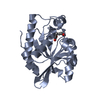
| ||||||||
| 2 | 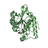
| ||||||||
| Unit cell |
|
-Components
| #1: Protein | Mass: 20832.793 Da / Num. of mol.: 2 Source method: isolated from a genetically manipulated source Source: (gene. exp.) Pseudomonas aeruginosa (bacteria) / Strain: ATCC 15692 / PAO1 / 1C / PRS 101 / LMG 12228 / Gene: PA4672, pth / Plasmid: pET-NH6 / Production host: #2: Chemical |   Mass: 160.168 Da / Num. of mol.: 2 / Source method: obtained synthetically / Formula: C7H12O4 Mass: 160.168 Da / Num. of mol.: 2 / Source method: obtained synthetically / Formula: C7H12O4#3: Water | ChemComp-HOH / |  Mass: 18.015 Da / Num. of mol.: 111 / Source method: isolated from a natural source / Formula: H2O Mass: 18.015 Da / Num. of mol.: 111 / Source method: isolated from a natural source / Formula: H2O |
|---|
-Experimental details
-Experiment
| Experiment | Method: X-RAY DIFFRACTION / Number of used crystals: 1 |
|---|
- Sample preparation
| Crystal | Density Matthews: 2.35 Å3/Da / Density % sol: 47.57 % |
|---|---|
| Crystal grow | Temperature: 298 K / Method: vapor diffusion, hanging drop / pH: 8.5 Details: 0.1M HEPES pH 8.5, PEG 4000, 5% Isopropanol, VAPOR DIFFUSION, HANGING DROP, temperature 298K |
-Data collection
| Diffraction | Mean temperature: 298 K |
|---|---|
| Diffraction source | Source: ROTATING ANODE / Type: RIGAKU RU300 / Wavelength: 1.514 Å |
| Detector | Type: MARRESEARCH / Detector: IMAGE PLATE / Date: Jan 1, 2012 / Details: Mirror |
| Radiation | Monochromator: Graphite / Protocol: SINGLE WAVELENGTH / Monochromatic (M) / Laue (L): M / Scattering type: x-ray |
| Radiation wavelength | Wavelength: 1.514 Å / Relative weight: 1 |
| Reflection | Resolution: 2.94→56.3 Å / Num. obs: 8159 / % possible obs: 99.5 % / Observed criterion σ(F): 0 / Observed criterion σ(I): 0 / Biso Wilson estimate: 53.7 Å2 / Rsym value: 0.147 / Net I/σ(I): 8.1 |
| Reflection shell | Resolution: 2.94→3.05 Å / Mean I/σ(I) obs: 2 / Rsym value: 0.393 / % possible all: 100 |
- Processing
| Software |
| ||||||||||||||||||||
|---|---|---|---|---|---|---|---|---|---|---|---|---|---|---|---|---|---|---|---|---|---|
| Refinement | Method to determine structure: MOLECULAR REPLACEMENT Starting model: PDB ENTRY 3P2J Resolution: 2.94→56.3 Å / Occupancy max: 1 / Occupancy min: 0.8 / Cross valid method: THROUGHOUT / σ(F): 0 / σ(I): 0 / Stereochemistry target values: Engh & Huber
| ||||||||||||||||||||
| Solvent computation | Bsol: 49.643 Å2 | ||||||||||||||||||||
| Displacement parameters | Biso max: 74.21 Å2 / Biso mean: 38.4112 Å2 / Biso min: 5.19 Å2
| ||||||||||||||||||||
| Refinement step | Cycle: LAST / Resolution: 2.94→56.3 Å
| ||||||||||||||||||||
| Refine LS restraints |
| ||||||||||||||||||||
| LS refinement shell | Resolution: 2.94→3.04 Å | ||||||||||||||||||||
| Xplor file |
|
