 Movie
Movie Controller

[English] 日本語
 Yorodumi
- PDB-3p2j: Crystal structure of peptidyl-tRNA hydrolase from Mycobacterium s... -
+ Open data

- Basic information
| Entry | Database: PDB / ID: 3p2j | ||||||
|---|---|---|---|---|---|---|---|
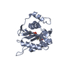
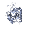
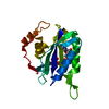
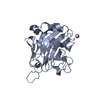
| Title | Crystal structure of peptidyl-tRNA hydrolase from Mycobacterium smegmatis at 2.2 A resolution | ||||||
 Components Components | Peptidyl-tRNA hydrolase | ||||||
 Keywords Keywords | HYDROLASE | ||||||
| Function / homology |  Function and homology information Function and homology informationpeptidyl-tRNA hydrolase / peptidyl-tRNA hydrolase activity / protein quality control for misfolded or incompletely synthesized proteins / rescue of stalled cytosolic ribosome / tRNA binding / cytoplasm Similarity search - Function | ||||||
| Biological species |  Mycobacterium smegmatis (bacteria) Mycobacterium smegmatis (bacteria) | ||||||
| Method |  X-RAY DIFFRACTION / MOLECULAR REPLACEMENT / Resolution: 2.22 Å X-RAY DIFFRACTION / MOLECULAR REPLACEMENT / Resolution: 2.22 Å | ||||||
 Authors Authors | Kumar, A. / Singh, A. / Yadav, R. / Sinha, M. / Arora, A. / Sharma, S. / Singh, T.P. | ||||||
 Citation Citation | Journal: To be Published Title: Crystal Structure of peptidyl-tRNA hydrolase from Mycobacterium smegmatis at 2.2 A resolution Authors: Kumar, A. / Singh, A. / Yadav, R. / Sinha, M. / Arora, A. / Sharma, S. / Singh, T.P. | ||||||
| History |
|
- Structure visualization
| Structure viewer | Molecule: MolmilJmol/JSmol |
|---|
- Downloads & links
-Download
| PDBx/mmCIF format | 3p2j.cif.gz | 52.2 KB | Display | PDBx/mmCIF format |
|---|---|---|---|---|
| PDB format | pdb3p2j.ent.gz | 36.4 KB | Display | PDB format |
| PDBx/mmJSON format | 3p2j.json.gz | Tree view | PDBx/mmJSON format | |
| Others |  Other downloads Other downloads |
-Validation report
| Arichive directory | https://data.pdbj.org/pub/pdb/validation_reports/p2/3p2jftp://data.pdbj.org/pub/pdb/validation_reports/p2/3p2j | HTTPS FTP |
|---|
-Related structure data
| Related structure data |  3kjzS S: Starting model for refinement |
|---|---|
| Similar structure data |


-Links
 PDBj
PDBj- Assembly
| Deposited unit | 
| ||||||||
|---|---|---|---|---|---|---|---|---|---|
| 1 |
| ||||||||
| Unit cell |
|
-Components
| #1: Protein | Mass: 20270.268 Da / Num. of mol.: 1 Source method: isolated from a genetically manipulated source Source: (gene. exp.) Mycobacterium smegmatis (bacteria) / Strain: ATCC 700084 / MC(2)155 / Production host: |
|---|---|
| #2: Water | ChemComp-HOH /  Mass: 18.015 Da / Num. of mol.: 155 / Source method: isolated from a natural source / Formula: H2O Mass: 18.015 Da / Num. of mol.: 155 / Source method: isolated from a natural source / Formula: H2O |
| Sequence details | AUTHOR STATED THAT THERE WERE ERRORS IN THE SEQUENCE DATABASE, THE SEQUENCE IN THIS ENTRY ...AUTHOR STATED THAT THERE WERE ERRORS IN THE SEQUENCE DATABASE, THE SEQUENCE IN THIS ENTRY REPRESENTE |
-Experimental details
-Experiment
| Experiment | Method: X-RAY DIFFRACTION / Number of used crystals: 1 |
|---|
- Sample preparation
| Crystal | Density Matthews: 2.02 Å3/Da / Density % sol: 39.09 % |
|---|---|
| Crystal grow | Temperature: 298 K / Method: vapor diffusion, hanging drop / pH: 7.5 Details: 20mM Tris-HCl, 1mM EDTA, 50mM NaCl, 10% isopropanol, 30% PEG 1500, 5mM 2-mercaptoethanol, pH 7.5, VAPOR DIFFUSION, HANGING DROP, temperature 298K |
-Data collection
| Diffraction | Mean temperature: 298 K |
|---|---|
| Diffraction source | Source: ROTATING ANODE / Type: RIGAKU RU300 / Wavelength: 1.514 Å |
| Detector | Type: MARRESEARCH / Detector: IMAGE PLATE / Date: May 26, 2009 / Details: MIRROR |
| Radiation | Monochromator: GRAPHITE / Protocol: SINGLE WAVELENGTH / Monochromatic (M) / Laue (L): M / Scattering type: x-ray |
| Radiation wavelength | Wavelength: 1.514 Å / Relative weight: 1 |
| Reflection | Resolution: 2.22→42.67 Å / Num. all: 8170 / Num. obs: 8170 / % possible obs: 95.6 % / Observed criterion σ(F): 0 / Observed criterion σ(I): 0 / Biso Wilson estimate: 32.3 Å2 / Rsym value: 0.078 / Net I/σ(I): 11.9 |
| Reflection shell | Resolution: 2.22→2.3 Å / Mean I/σ(I) obs: 2 / Rsym value: 0.641 / % possible all: 89.4 |
- Processing
| Software |
| |||||||||||||||||||||||||||||||||||||||||||||||||||||||||||||||||
|---|---|---|---|---|---|---|---|---|---|---|---|---|---|---|---|---|---|---|---|---|---|---|---|---|---|---|---|---|---|---|---|---|---|---|---|---|---|---|---|---|---|---|---|---|---|---|---|---|---|---|---|---|---|---|---|---|---|---|---|---|---|---|---|---|---|---|
| Refinement | Method to determine structure: MOLECULAR REPLACEMENT Starting model: PDB ENTRY 3KJZ Resolution: 2.22→14.96 Å / Cor.coef. Fo:Fc: 0.958 / Cor.coef. Fo:Fc free: 0.94 / Isotropic thermal model: RESTRAINED / Cross valid method: THROUGHOUT / σ(F): 0 / σ(I): 0 / ESU R Free: 0.226 / Stereochemistry target values: Engh & Huber / Details: HYDROGENS HAVE BEEN ADDED IN THE RIDING POSITIONS
| |||||||||||||||||||||||||||||||||||||||||||||||||||||||||||||||||
| Solvent computation | Ion probe radii: 0.8 Å / Shrinkage radii: 0.8 Å / VDW probe radii: 1.4 Å / Solvent model: MASK | |||||||||||||||||||||||||||||||||||||||||||||||||||||||||||||||||
| Displacement parameters | Biso mean: 36.27 Å2
| |||||||||||||||||||||||||||||||||||||||||||||||||||||||||||||||||
| Refine analyze |
| |||||||||||||||||||||||||||||||||||||||||||||||||||||||||||||||||
| Refinement step | Cycle: LAST / Resolution: 2.22→14.96 Å
| |||||||||||||||||||||||||||||||||||||||||||||||||||||||||||||||||
| Refine LS restraints |
| |||||||||||||||||||||||||||||||||||||||||||||||||||||||||||||||||
| LS refinement shell | Resolution: 2.224→2.282 Å / Total num. of bins used: 20
|
