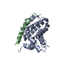
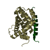
Deposited unit
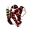
A: Induced myeloid leukemia cell differentiation protein Mcl-1
B: Mule BH3 peptide from E3 ubiquitin-protein ligase HUWE1
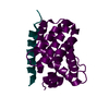
C: Induced myeloid leukemia cell differentiation protein Mcl-1
D: Mule BH3 peptide from E3 ubiquitin-protein ligase HUWE1
E: Induced myeloid leukemia cell differentiation protein Mcl-1
F: Mule BH3 peptide from E3 ubiquitin-protein ligase HUWE1
G: Induced myeloid leukemia cell differentiation protein Mcl-1
H: Mule BH3 peptide from E3 ubiquitin-protein ligase HUWE1
I: Induced myeloid leukemia cell differentiation protein Mcl-1
J: Mule BH3 peptide from E3 ubiquitin-protein ligase HUWE1
K: Induced myeloid leukemia cell differentiation protein Mcl-1
L: Mule BH3 peptide from E3 ubiquitin-protein ligase HUWE1
M: Induced myeloid leukemia cell differentiation protein Mcl-1
N: Mule BH3 peptide from E3 ubiquitin-protein ligase HUWE1
O: Induced myeloid leukemia cell differentiation protein Mcl-1
P: Mule BH3 peptide from E3 ubiquitin-protein ligase HUWE1
Q: Induced myeloid leukemia cell differentiation protein Mcl-1
R: Mule BH3 peptide from E3 ubiquitin-protein ligase HUWE1
S: Induced myeloid leukemia cell differentiation protein Mcl-1
T: Mule BH3 peptide from E3 ubiquitin-protein ligase HUWE1
U: Induced myeloid leukemia cell differentiation protein Mcl-1
V: Mule BH3 peptide from E3 ubiquitin-protein ligase HUWE1
W: Induced myeloid leukemia cell differentiation protein Mcl-1
X: Mule BH3 peptide from E3 ubiquitin-protein ligase HUWE1 Summary Component details
Theoretical mass Number of molelcules Total (without water) 250,736 24 Polymers 250,736 24 Non-polymers 0 0 Water 3,099 172
1 Summary Symmetry operations Calculated values
Idetical with deposited unit defined by author
Type Name Symmetry operation Number identity operation 1_555 x,y,z 1
Buried area 39980 Å2 ΔGint -208 kcal/mol Surface area 96530 Å2
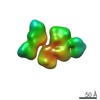
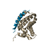
2
A: Induced myeloid leukemia cell differentiation protein Mcl-1
B: Mule BH3 peptide from E3 ubiquitin-protein ligase HUWE1 Summary Component details Symmetry operations Calculated values
Theoretical mass Number of molelcules Total (without water) 20,895 2 Polymers 20,895 2 Non-polymers 0 0 Water 36 2
Type Name Symmetry operation Number identity operation 1_555 x,y,z 1
Buried area 2050 Å2 ΔGint -17 kcal/mol Surface area 9400 Å2 Method
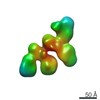
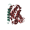
3
C: Induced myeloid leukemia cell differentiation protein Mcl-1
D: Mule BH3 peptide from E3 ubiquitin-protein ligase HUWE1 Summary Component details Symmetry operations Calculated values
Theoretical mass Number of molelcules Total (without water) 20,895 2 Polymers 20,895 2 Non-polymers 0 0 Water 36 2
Type Name Symmetry operation Number identity operation 1_555 x,y,z 1
Buried area 1890 Å2 ΔGint -18 kcal/mol Surface area 9660 Å2 Method
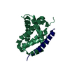
4
E: Induced myeloid leukemia cell differentiation protein Mcl-1
F: Mule BH3 peptide from E3 ubiquitin-protein ligase HUWE1 Summary Component details Symmetry operations Calculated values
Theoretical mass Number of molelcules Total (without water) 20,895 2 Polymers 20,895 2 Non-polymers 0 0 Water 18 1
Type Name Symmetry operation Number identity operation 1_555 x,y,z 1
Buried area 2040 Å2 ΔGint -14 kcal/mol Surface area 9520 Å2 Method
5
G: Induced myeloid leukemia cell differentiation protein Mcl-1
H: Mule BH3 peptide from E3 ubiquitin-protein ligase HUWE1 Summary Component details Symmetry operations Calculated values
Theoretical mass Number of molelcules Total (without water) 20,895 2 Polymers 20,895 2 Non-polymers 0 0 Water 36 2
Type Name Symmetry operation Number identity operation 1_555 x,y,z 1
Buried area 1960 Å2 ΔGint -17 kcal/mol Surface area 9380 Å2 Method
6
I: Induced myeloid leukemia cell differentiation protein Mcl-1
J: Mule BH3 peptide from E3 ubiquitin-protein ligase HUWE1 Summary Component details Symmetry operations Calculated values
Theoretical mass Number of molelcules Total (without water) 20,895 2 Polymers 20,895 2 Non-polymers 0 0 Water 36 2
Type Name Symmetry operation Number identity operation 1_555 x,y,z 1
Buried area 2010 Å2 ΔGint -17 kcal/mol Surface area 9500 Å2 Method
7
K: Induced myeloid leukemia cell differentiation protein Mcl-1
L: Mule BH3 peptide from E3 ubiquitin-protein ligase HUWE1 Summary Component details Symmetry operations Calculated values
Theoretical mass Number of molelcules Total (without water) 20,895 2 Polymers 20,895 2 Non-polymers 0 0 Water 36 2
Type Name Symmetry operation Number identity operation 1_555 x,y,z 1
Buried area 1950 Å2 ΔGint -18 kcal/mol Surface area 9560 Å2 Method
8
M: Induced myeloid leukemia cell differentiation protein Mcl-1
N: Mule BH3 peptide from E3 ubiquitin-protein ligase HUWE1 Summary Component details Symmetry operations Calculated values
Theoretical mass Number of molelcules Total (without water) 20,895 2 Polymers 20,895 2 Non-polymers 0 0 Water 36 2
Type Name Symmetry operation Number identity operation 1_555 x,y,z 1
Buried area 2060 Å2 ΔGint -17 kcal/mol Surface area 9650 Å2 Method
9
O: Induced myeloid leukemia cell differentiation protein Mcl-1
P: Mule BH3 peptide from E3 ubiquitin-protein ligase HUWE1 Summary Component details Symmetry operations Calculated values
Theoretical mass Number of molelcules Total (without water) 20,895 2 Polymers 20,895 2 Non-polymers 0 0 Water 36 2
Type Name Symmetry operation Number identity operation 1_555 x,y,z 1
Buried area 1890 Å2 ΔGint -19 kcal/mol Surface area 9530 Å2 Method
10
Q: Induced myeloid leukemia cell differentiation protein Mcl-1
R: Mule BH3 peptide from E3 ubiquitin-protein ligase HUWE1 Summary Component details Symmetry operations Calculated values
Theoretical mass Number of molelcules Total (without water) 20,895 2 Polymers 20,895 2 Non-polymers 0 0 Water 36 2
Type Name Symmetry operation Number identity operation 1_555 x,y,z 1
Buried area 2030 Å2 ΔGint -18 kcal/mol Surface area 9200 Å2 Method
11
S: Induced myeloid leukemia cell differentiation protein Mcl-1
T: Mule BH3 peptide from E3 ubiquitin-protein ligase HUWE1 Summary Component details Symmetry operations Calculated values
Theoretical mass Number of molelcules Total (without water) 20,895 2 Polymers 20,895 2 Non-polymers 0 0 Water 36 2
Type Name Symmetry operation Number identity operation 1_555 x,y,z 1
Buried area 1870 Å2 ΔGint -18 kcal/mol Surface area 9400 Å2 Method
12
U: Induced myeloid leukemia cell differentiation protein Mcl-1
V: Mule BH3 peptide from E3 ubiquitin-protein ligase HUWE1 Summary Component details Symmetry operations Calculated values
Theoretical mass Number of molelcules Total (without water) 20,895 2 Polymers 20,895 2 Non-polymers 0 0 Water 36 2
Type Name Symmetry operation Number identity operation 1_555 x,y,z 1
Buried area 1940 Å2 ΔGint -18 kcal/mol Surface area 8820 Å2 Method
13
W: Induced myeloid leukemia cell differentiation protein Mcl-1
X: Mule BH3 peptide from E3 ubiquitin-protein ligase HUWE1 Summary Component details Symmetry operations Calculated values
Theoretical mass Number of molelcules Total (without water) 20,895 2 Polymers 20,895 2 Non-polymers 0 0 Water 36 2
Type Name Symmetry operation Number identity operation 1_555 x,y,z 1
Buried area 1940 Å2 ΔGint -18 kcal/mol Surface area 9260 Å2 Method
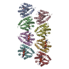
Unit cell Length a, b, c (Å) 31.790, 114.240, 135.980 Angle α, β, γ (deg.) 90.12, 92.32, 90.01 Int Tables number 1 Space group name H-M P1
 Movie
Movie Controller


 Open data
 Basic information
 Components
 Keywords
 Function and homology information
 Homo sapiens (human)
 X-RAY DIFFRACTION /
  Authors
 China, 2items
  Citation
 Structure visualization
 Downloads & links
 Other downloads
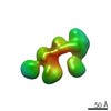

 PDBj
PDBj


 Assembly


 Mass: 18.015 Da / Num. of mol.: 172 / Source method: isolated from a natural source / Formula: H2O
 Sample preparation
 / Beamline: 8.3.1 / Wavelength: 0.987, 0.988, 1.0
 Processing