 Movie
Movie Controller

[English] 日本語
 Yorodumi
- PDB-4fop: Crystal Structure of Peptidyl-tRNA hydrolase from Acinetobacter b... -
+ Open data

- Basic information
| Entry | Database: PDB / ID: 4fop | ||||||
|---|---|---|---|---|---|---|---|
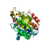
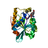
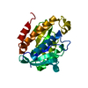
| Title | Crystal Structure of Peptidyl-tRNA hydrolase from Acinetobacter baumannii at 1.86 A resolution | ||||||
 Components Components | Peptidyl-tRNA hydrolase | ||||||
 Keywords Keywords | HYDROLASE | ||||||
| Function / homology |  Function and homology information Function and homology informationpeptidyl-tRNA hydrolase / peptidyl-tRNA hydrolase activity / protein quality control for misfolded or incompletely synthesized proteins / rescue of stalled cytosolic ribosome / tRNA binding / cytoplasm Similarity search - Function | ||||||
| Biological species |  Acinetobacter baumannii (bacteria) Acinetobacter baumannii (bacteria) | ||||||
| Method |  X-RAY DIFFRACTION / SYNCHROTRON / MOLECULAR REPLACEMENT / Resolution: 1.86 Å X-RAY DIFFRACTION / SYNCHROTRON / MOLECULAR REPLACEMENT / Resolution: 1.86 Å | ||||||
 Authors Authors | Kaushik, S. / Kumar, S. / Singh, N. / Sinha, M. / Kaur, P. / Sharma, S. / Singh, T.P. | ||||||
 Citation Citation | Journal: Plos One / Year: 2013 Title: The Mode of Inhibitor Binding to Peptidyl-tRNA Hydrolase: Binding Studies and Structure Determination of Unbound and Bound Peptidyl-tRNA Hydrolase from Acinetobacter baumannii Authors: Kaushik, S. / Singh, N. / Yamini, S. / Singh, A. / Sinha, M. / Arora, A. / Kaur, P. / Sharma, S. / Singh, T.P. | ||||||
| History |
|
- Structure visualization
| Structure viewer | Molecule: MolmilJmol/JSmol |
|---|
- Downloads & links
-Download
| PDBx/mmCIF format | 4fop.cif.gz | 55.8 KB | Display | PDBx/mmCIF format |
|---|---|---|---|---|
| PDB format | pdb4fop.ent.gz | 39.5 KB | Display | PDB format |
| PDBx/mmJSON format | 4fop.json.gz | Tree view | PDBx/mmJSON format | |
| Others |  Other downloads Other downloads |
-Validation report
| Arichive directory | https://data.pdbj.org/pub/pdb/validation_reports/fo/4fopftp://data.pdbj.org/pub/pdb/validation_reports/fo/4fop | HTTPS FTP |
|---|
-Related structure data
| Related structure data | 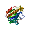 4fotC 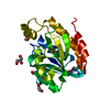 4hoyC  4ikoC 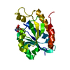 4jwkC  4jx9C 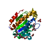 4jy7C  2pthS S: Starting model for refinement C: citing same article ( |
|---|---|
| Similar structure data |

-Links
 PDBj
PDBj- Assembly
| Deposited unit | 
| ||||||||
|---|---|---|---|---|---|---|---|---|---|
| 1 |
| ||||||||
| Unit cell |
| ||||||||
| Components on special symmetry positions |
|
-Components
| #1: Protein | Mass: 20967.957 Da / Num. of mol.: 1 Source method: isolated from a genetically manipulated source Source: (gene. exp.) Acinetobacter baumannii (bacteria) / Strain: ATCC19606 / Gene: pth / Plasmid: pET28a / Production host: | ||||||
|---|---|---|---|---|---|---|---|
| #2: Chemical |   Mass: 92.094 Da / Num. of mol.: 2 / Source method: obtained synthetically / Formula: C3H8O3 Mass: 92.094 Da / Num. of mol.: 2 / Source method: obtained synthetically / Formula: C3H8O3#3: Chemical | ChemComp-PEG /   Mass: 106.120 Da / Num. of mol.: 4 / Source method: obtained synthetically / Formula: C4H10O3 Mass: 106.120 Da / Num. of mol.: 4 / Source method: obtained synthetically / Formula: C4H10O3#4: Chemical | ChemComp-ACT / |   Mass: 59.044 Da / Num. of mol.: 1 / Source method: obtained synthetically / Formula: C2H3O2 Mass: 59.044 Da / Num. of mol.: 1 / Source method: obtained synthetically / Formula: C2H3O2#5: Water | ChemComp-HOH / |  Mass: 18.015 Da / Num. of mol.: 179 / Source method: isolated from a natural source / Formula: H2O Mass: 18.015 Da / Num. of mol.: 179 / Source method: isolated from a natural source / Formula: H2O |
-Experimental details
-Experiment
| Experiment | Method: X-RAY DIFFRACTION / Number of used crystals: 1 |
|---|
- Sample preparation
| Crystal | Density Matthews: 2.59 Å3/Da / Density % sol: 52.57 % |
|---|---|
| Crystal grow | Temperature: 310 K / Method: vapor diffusion, hanging drop / pH: 6.5 Details: 0.2M HEPES Buffer, 25% PEG10000, pH 6.5, VAPOR DIFFUSION, HANGING DROP, temperature 310K |
-Data collection
| Diffraction | Mean temperature: 77 K |
|---|---|
| Diffraction source | Source: SYNCHROTRON / Site: ESRF  / Beamline: BM14 / Wavelength: 0.97 Å / Beamline: BM14 / Wavelength: 0.97 Å |
| Detector | Type: MARRESEARCH / Detector: CCD / Date: May 18, 2012 / Details: MIRROR |
| Radiation | Monochromator: Graphite / Protocol: SINGLE WAVELENGTH / Monochromatic (M) / Laue (L): M / Scattering type: x-ray |
| Radiation wavelength | Wavelength: 0.97 Å / Relative weight: 1 |
| Reflection | Resolution: 1.86→57.8 Å / Num. all: 18987 / Num. obs: 17986 / % possible obs: 99.1 % / Observed criterion σ(F): 0 / Observed criterion σ(I): 0 / Rsym value: 0.062 / Net I/σ(I): 11.6 |
| Reflection shell | Resolution: 1.86→1.93 Å / Mean I/σ(I) obs: 1.7 / Num. unique all: 18987 / Rsym value: 0.573 / % possible all: 99.4 |
- Processing
| Software |
| |||||||||||||||||||||||||||||||||||||||||||||
|---|---|---|---|---|---|---|---|---|---|---|---|---|---|---|---|---|---|---|---|---|---|---|---|---|---|---|---|---|---|---|---|---|---|---|---|---|---|---|---|---|---|---|---|---|---|---|
| Refinement | Method to determine structure: MOLECULAR REPLACEMENT Starting model: 2PTH Resolution: 1.86→57.8 Å / Cor.coef. Fo:Fc: 0.964 / Cor.coef. Fo:Fc free: 0.937 / SU B: 3.095 / SU ML: 0.092 / Cross valid method: THROUGHOUT / σ(F): 0 / σ(I): 0 / ESU R: 0.129 / ESU R Free: 0.133 / Stereochemistry target values: MAXIMUM LIKELIHOOD / Details: HYDROGENS HAVE BEEN USED IF PRESENT IN THE INPUT
| |||||||||||||||||||||||||||||||||||||||||||||
| Solvent computation | Ion probe radii: 0.8 Å / Shrinkage radii: 0.8 Å / VDW probe radii: 1.2 Å / Solvent model: MASK | |||||||||||||||||||||||||||||||||||||||||||||
| Displacement parameters | Biso mean: 27.029 Å2
| |||||||||||||||||||||||||||||||||||||||||||||
| Refinement step | Cycle: LAST / Resolution: 1.86→57.8 Å
| |||||||||||||||||||||||||||||||||||||||||||||
| Refine LS restraints |
| |||||||||||||||||||||||||||||||||||||||||||||
| LS refinement shell | Resolution: 1.86→1.93 Å / Total num. of bins used: 20
|
