 Movie
Movie Controller

[English] 日本語
 Yorodumi
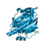
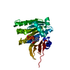
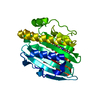
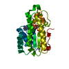
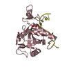
- PDB-6ubc: Crystal structure of a GH128 (subgroup VII) oligosaccharide-bindi... -
+ Open data

- Basic information
| Entry | Database: PDB / ID: 6ubc | ||||||
|---|---|---|---|---|---|---|---|
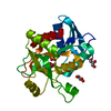
| Title | Crystal structure of a GH128 (subgroup VII) oligosaccharide-binding protein from Cryptococcus neoformans (CnGH128_VII) | ||||||
 Components Components | Glyco_hydro_cc domain-containing protein | ||||||
 Keywords Keywords | HYDROLASE / Glycosyl hydrolase / CARBOHYDRATE | ||||||
| Function / homology | : / Uncharacterised protein family, glycosyl hydrolase catalytic domain / Glycosyl hydrolase catalytic core / Glycoside hydrolase superfamily / Asl1-like glycosyl hydrolase catalytic domain-containing protein Function and homology information Function and homology information | ||||||
| Biological species |  Cryptococcus neoformans (Cryptococcus neoformans serotype A) Cryptococcus neoformans (Cryptococcus neoformans serotype A) | ||||||
| Method |  X-RAY DIFFRACTION / SYNCHROTRON / MOLECULAR REPLACEMENT / Resolution: 1.65 Å X-RAY DIFFRACTION / SYNCHROTRON / MOLECULAR REPLACEMENT / Resolution: 1.65 Å | ||||||
 Authors Authors | Santos, C.R. / Costa, P.A.C.R. / Souza, B.P. / Vieira, P.S. / Murakami, M.T. | ||||||
| Funding support |  Brazil, 1items Brazil, 1items
| ||||||
 Citation Citation | Journal: Nat.Chem.Biol. / Year: 2020 Title: Structural insights into beta-1,3-glucan cleavage by a glycoside hydrolase family. Authors: Santos, C.R. / Costa, P.A.C.R. / Vieira, P.S. / Gonzalez, S.E.T. / Correa, T.L.R. / Lima, E.A. / Mandelli, F. / Pirolla, R.A.S. / Domingues, M.N. / Cabral, L. / Martins, M.P. / Cordeiro, R.L. ...Authors: Santos, C.R. / Costa, P.A.C.R. / Vieira, P.S. / Gonzalez, S.E.T. / Correa, T.L.R. / Lima, E.A. / Mandelli, F. / Pirolla, R.A.S. / Domingues, M.N. / Cabral, L. / Martins, M.P. / Cordeiro, R.L. / Junior, A.T. / Souza, B.P. / Prates, E.T. / Gozzo, F.C. / Persinoti, G.F. / Skaf, M.S. / Murakami, M.T. | ||||||
| History |
|
- Structure visualization
| Structure viewer | Molecule: MolmilJmol/JSmol |
|---|
- Downloads & links
-Download
| PDBx/mmCIF format | 6ubc.cif.gz | 73.4 KB | Display | PDBx/mmCIF format |
|---|---|---|---|---|
| PDB format | pdb6ubc.ent.gz | 51.2 KB | Display | PDB format |
| PDBx/mmJSON format | 6ubc.json.gz | Tree view | PDBx/mmJSON format | |
| Others |  Other downloads Other downloads |
-Validation report
| Arichive directory | https://data.pdbj.org/pub/pdb/validation_reports/ub/6ubcftp://data.pdbj.org/pub/pdb/validation_reports/ub/6ubc | HTTPS FTP |
|---|
-Related structure data
| Related structure data | 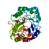 6uaqC  6uarC 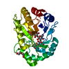 6uasC  6uatC 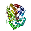 6uauC 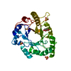 6uavC 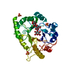 6uawC  6uaxC 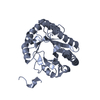 6uayC 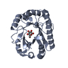 6uazC 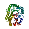 6ub0C 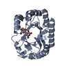 6ub1C 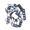 6ub2C 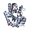 6ub3C 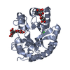 6ub4C 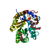 6ub5C 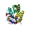 6ub6C 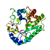 6ub7C 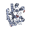 6ub8C 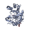 6ubaC 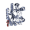 6ubbC 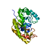 6ubdC 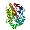 6uflC 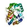 6ufzC C: citing same article ( |
|---|---|
| Similar structure data |


-Links
 PDBj
PDBj- Assembly
| Deposited unit | 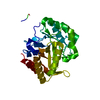
| ||||||||
|---|---|---|---|---|---|---|---|---|---|
| 1 |
| ||||||||
| Unit cell |
| ||||||||
| Components on special symmetry positions |
|
-Components
| #1: Protein | Mass: 46711.031 Da / Num. of mol.: 1 Source method: isolated from a genetically manipulated source Source: (gene. exp.) Cryptococcus neoformans (Cryptococcus neoformans serotype A)Production host:  |
|---|---|
| #2: Water | ChemComp-HOH /  Mass: 18.015 Da / Num. of mol.: 298 / Source method: isolated from a natural source / Formula: H2O Mass: 18.015 Da / Num. of mol.: 298 / Source method: isolated from a natural source / Formula: H2O |
| Has protein modification | Y |
-Experimental details
-Experiment
| Experiment | Method: X-RAY DIFFRACTION / Number of used crystals: 1 |
|---|
- Sample preparation
| Crystal | Density Matthews: 1.48 Å3/Da / Density % sol: 16.9 % |
|---|---|
| Crystal grow | Temperature: 291 K / Method: vapor diffusion, sitting drop / pH: 4.6 / Details: Sodium acetate 0.1 M ammonium sulfate 2.0 M |
-Data collection
| Diffraction | Mean temperature: 100 K / Serial crystal experiment: N |
|---|---|
| Diffraction source | Source: SYNCHROTRON / Site: SSRL  / Beamline: BL12-2 / Wavelength: 0.97946 Å / Beamline: BL12-2 / Wavelength: 0.97946 Å |
| Detector | Type: DECTRIS PILATUS 6M / Detector: PIXEL / Date: May 18, 2018 |
| Radiation | Protocol: SINGLE WAVELENGTH / Monochromatic (M) / Laue (L): M / Scattering type: x-ray |
| Radiation wavelength | Wavelength: 0.97946 Å / Relative weight: 1 |
| Reflection | Resolution: 1.65→48.39 Å / Num. obs: 33192 / % possible obs: 99.8 % / Redundancy: 40.1 % / CC1/2: 0.999 / Net I/σ(I): 16.65 |
| Reflection shell | Resolution: 1.65→1.75 Å / Num. unique obs: 5316 / CC1/2: 0.589 |
- Processing
| Software |
| |||||||||||||||||||||||||||||||||||||||||||||||||||||||||||||||||||||||||||
|---|---|---|---|---|---|---|---|---|---|---|---|---|---|---|---|---|---|---|---|---|---|---|---|---|---|---|---|---|---|---|---|---|---|---|---|---|---|---|---|---|---|---|---|---|---|---|---|---|---|---|---|---|---|---|---|---|---|---|---|---|---|---|---|---|---|---|---|---|---|---|---|---|---|---|---|---|
| Refinement | Method to determine structure: MOLECULAR REPLACEMENT / Resolution: 1.65→48.39 Å / Cor.coef. Fo:Fc: 0.976 / Cor.coef. Fo:Fc free: 0.959 / Cross valid method: THROUGHOUT / σ(F): 0 / ESU R: 0.09 / ESU R Free: 0.093 Details: HYDROGENS HAVE BEEN ADDED IN THE RIDING POSITIONS U VALUES : REFINED INDIVIDUALLY
| |||||||||||||||||||||||||||||||||||||||||||||||||||||||||||||||||||||||||||
| Solvent computation | Ion probe radii: 0.8 Å / Shrinkage radii: 0.8 Å / VDW probe radii: 1.2 Å | |||||||||||||||||||||||||||||||||||||||||||||||||||||||||||||||||||||||||||
| Displacement parameters | Biso max: 75.3 Å2 / Biso mean: 27.362 Å2 / Biso min: 19.98 Å2
| |||||||||||||||||||||||||||||||||||||||||||||||||||||||||||||||||||||||||||
| Refinement step | Cycle: final / Resolution: 1.65→48.39 Å
| |||||||||||||||||||||||||||||||||||||||||||||||||||||||||||||||||||||||||||
| Refine LS restraints |
| |||||||||||||||||||||||||||||||||||||||||||||||||||||||||||||||||||||||||||
| LS refinement shell | Resolution: 1.651→1.694 Å / Total num. of bins used: 20
|
