 Movie
Movie Controller


+ Open data

- Basic information
| Entry | Database: PDB / ID: 3i95 | ||||||
|---|---|---|---|---|---|---|---|
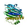
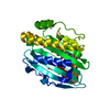
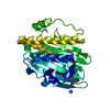
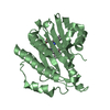
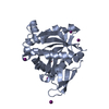
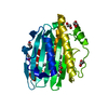
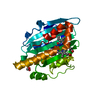
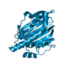
| Title | Crystal structure of E76Q mutant PcyA-biliverdin complex | ||||||
 Components Components | Phycocyanobilin:ferredoxin oxidoreductase | ||||||
 Keywords Keywords | OXIDOREDUCTASE / alpha-beta-alpha sandwich / mutant enzyme-substrate complex | ||||||
| Function / homology |  Function and homology information Function and homology informationphycocyanobilin:ferredoxin oxidoreductase / phycocyanobilin:ferredoxin oxidoreductase activity / phytochromobilin biosynthetic process / cobalt ion binding Similarity search - Function | ||||||
| Biological species |  | ||||||
| Method |  X-RAY DIFFRACTION / SYNCHROTRON / DIFFERENCE FOURIER / Resolution: 1.4 Å X-RAY DIFFRACTION / SYNCHROTRON / DIFFERENCE FOURIER / Resolution: 1.4 Å | ||||||
 Authors Authors | Hagiwara, Y. / Sugishima, M. / Fukuyama, K. | ||||||
 Citation Citation | Journal: To be Published Title: Structural insights into vinyl reduction regiospecificity of phycocyanobilin:ferredoxin oxidoreductase (PcyA). Authors: Hagiwara, Y. / Sugishima, M. / Khawn, H. / Kinoshita, H. / Inomata, K. / Shang, L. / Lagarias, J.C. / Takahashi, Y. / Fukuyama, K. | ||||||
| History |
|
- Structure visualization
| Structure viewer | Molecule: MolmilJmol/JSmol |
|---|
- Downloads & links
-Download
| PDBx/mmCIF format | 3i95.cif.gz | 74.8 KB | Display | PDBx/mmCIF format |
|---|---|---|---|---|
| PDB format | pdb3i95.ent.gz | 54.5 KB | Display | PDB format |
| PDBx/mmJSON format | 3i95.json.gz | Tree view | PDBx/mmJSON format | |
| Others |  Other downloads Other downloads |
-Validation report
| Arichive directory | https://data.pdbj.org/pub/pdb/validation_reports/i9/3i95ftp://data.pdbj.org/pub/pdb/validation_reports/i9/3i95 | HTTPS FTP |
|---|
-Related structure data
| Related structure data |  3i8uC 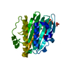 3i94C  2d1eS S: Starting model for refinement C: citing same article ( |
|---|---|
| Similar structure data |


-Links
 PDBj
PDBj- Assembly
| Deposited unit | 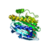
| ||||||||
|---|---|---|---|---|---|---|---|---|---|
| 1 |
| ||||||||
| Unit cell |
| ||||||||
| Components on special symmetry positions |
| ||||||||
| Details | biological unit is the same as asym. |
-Components
| #1: Protein | Mass: 28155.170 Da / Num. of mol.: 1 / Mutation: E76Q Source method: isolated from a genetically manipulated source Source: (gene. exp.) |
|---|---|
| #2: Chemical | ChemComp-BLA /   Mass: 582.646 Da / Num. of mol.: 1 / Source method: obtained synthetically / Formula: C33H34N4O6 Mass: 582.646 Da / Num. of mol.: 1 / Source method: obtained synthetically / Formula: C33H34N4O6 |
| #3: Water | ChemComp-HOH /  Mass: 18.015 Da / Num. of mol.: 334 / Source method: isolated from a natural source / Formula: H2O Mass: 18.015 Da / Num. of mol.: 334 / Source method: isolated from a natural source / Formula: H2O |
-Experimental details
-Experiment
| Experiment | Method: X-RAY DIFFRACTION / Number of used crystals: 1 |
|---|
- Sample preparation
| Crystal | Density Matthews: 2.57 Å3/Da / Density % sol: 52.07 % |
|---|---|
| Crystal grow | Temperature: 293 K / Method: vapor diffusion, hanging drop / pH: 7 Details: 1.7M ammonium sulfate, 2% PEG 400, 0.1M HEPES (pH 7.0), vapor diffusion, hanging drop, temperature 293K |
-Data collection
| Diffraction | Mean temperature: 100 K |
|---|---|
| Diffraction source | Source: SYNCHROTRON / Site: SPring-8  / Beamline: BL38B1 / Wavelength: 1 Å / Beamline: BL38B1 / Wavelength: 1 Å |
| Detector | Type: RIGAKU JUPITER 210 / Detector: CCD / Date: Feb 26, 2009 |
| Radiation | Monochromator: SI(111) DOUBLE MONOCHROMATER / Protocol: SINGLE WAVELENGTH / Monochromatic (M) / Laue (L): M / Scattering type: x-ray |
| Radiation wavelength | Wavelength: 1 Å / Relative weight: 1 |
| Reflection | Resolution: 1.4→17.98 Å / Num. obs: 57594 / % possible obs: 99.4 % / Redundancy: 6.7 % / Rmerge(I) obs: 0.055 |
| Reflection shell | Resolution: 1.4→1.45 Å / Redundancy: 6 % / Rmerge(I) obs: 0.295 / % possible all: 98.4 |
- Processing
| Software |
| ||||||||||||||||||||||||||||||||||||||||||||||||||||||||||||||||||||||||||||||||||||||||||
|---|---|---|---|---|---|---|---|---|---|---|---|---|---|---|---|---|---|---|---|---|---|---|---|---|---|---|---|---|---|---|---|---|---|---|---|---|---|---|---|---|---|---|---|---|---|---|---|---|---|---|---|---|---|---|---|---|---|---|---|---|---|---|---|---|---|---|---|---|---|---|---|---|---|---|---|---|---|---|---|---|---|---|---|---|---|---|---|---|---|---|---|
| Refinement | Method to determine structure: DIFFERENCE FOURIER Starting model: PDB ENTRY 2D1E Resolution: 1.4→17.98 Å / Cor.coef. Fo:Fc: 0.964 / Cor.coef. Fo:Fc free: 0.959 / Occupancy max: 1 / Occupancy min: 0.2 / SU B: 0.858 / SU ML: 0.035 / Cross valid method: THROUGHOUT / σ(F): 0 / ESU R: 0.058 / ESU R Free: 0.058 / Stereochemistry target values: MAXIMUM LIKELIHOOD / Details: HYDROGENS HAVE BEEN ADDED IN THE RIDING POSITIONS
| ||||||||||||||||||||||||||||||||||||||||||||||||||||||||||||||||||||||||||||||||||||||||||
| Solvent computation | Ion probe radii: 0.8 Å / Shrinkage radii: 0.8 Å / VDW probe radii: 1.4 Å / Solvent model: BABINET MODEL WITH MASK | ||||||||||||||||||||||||||||||||||||||||||||||||||||||||||||||||||||||||||||||||||||||||||
| Displacement parameters | Biso max: 60.44 Å2 / Biso mean: 16.171 Å2 / Biso min: 7.56 Å2
| ||||||||||||||||||||||||||||||||||||||||||||||||||||||||||||||||||||||||||||||||||||||||||
| Refinement step | Cycle: LAST / Resolution: 1.4→17.98 Å
| ||||||||||||||||||||||||||||||||||||||||||||||||||||||||||||||||||||||||||||||||||||||||||
| Refine LS restraints |
| ||||||||||||||||||||||||||||||||||||||||||||||||||||||||||||||||||||||||||||||||||||||||||
| LS refinement shell | Resolution: 1.4→1.436 Å / Total num. of bins used: 20
|
