 Movie
Movie Controller


+ Open data

- Basic information
| Entry | Database: PDB / ID: 3f0m | ||||||
|---|---|---|---|---|---|---|---|
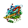
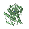
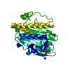
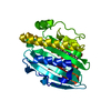
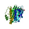
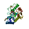
| Title | Crystal structure of radical D105N Synechocystis sp. PcyA | ||||||
 Components Components | Phycocyanobilin:ferredoxin oxidoreductase | ||||||
 Keywords Keywords | OXIDOREDUCTASE / ALPHA-BETA-ALPHA SANDWICH / RADICAL CHEMISTRY | ||||||
| Function / homology |  Function and homology information Function and homology informationphycocyanobilin:ferredoxin oxidoreductase / phycocyanobilin:ferredoxin oxidoreductase activity / phytochromobilin biosynthetic process / cobalt ion binding Similarity search - Function | ||||||
| Biological species |  | ||||||
| Method |  X-RAY DIFFRACTION / SYNCHROTRON / MOLECULAR REPLACEMENT / molecular replacement / Resolution: 1.5 Å X-RAY DIFFRACTION / SYNCHROTRON / MOLECULAR REPLACEMENT / molecular replacement / Resolution: 1.5 Å | ||||||
 Authors Authors | Kohler, A.C. / Gae, D.D. | ||||||
 Citation Citation | Journal: Biochemistry / Year: 2010 Title: Structural basis for hydration dynamics in radical stabilization of bilin reductase mutants. Authors: Kohler, A.C. / Gae, D.D. / Richley, M.A. / Stoll, S. / Gunn, A. / Lim, S. / Martin, S.S. / Doukov, T.I. / Britt, R.D. / Ames, J.B. / Lagarias, J.C. / Fisher, A.J. | ||||||
| History |
|
- Structure visualization
| Structure viewer | Molecule: MolmilJmol/JSmol |
|---|
- Downloads & links
-Download
| PDBx/mmCIF format | 3f0m.cif.gz | 72.7 KB | Display | PDBx/mmCIF format |
|---|---|---|---|---|
| PDB format | pdb3f0m.ent.gz | 53.3 KB | Display | PDB format |
| PDBx/mmJSON format | 3f0m.json.gz | Tree view | PDBx/mmJSON format | |
| Others |  Other downloads Other downloads |
-Validation report
| Arichive directory | https://data.pdbj.org/pub/pdb/validation_reports/f0/3f0mftp://data.pdbj.org/pub/pdb/validation_reports/f0/3f0m | HTTPS FTP |
|---|
-Related structure data


-Links
 PDBj
PDBj- Assembly
| Deposited unit | 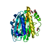
| ||||||||
|---|---|---|---|---|---|---|---|---|---|
| 1 |
| ||||||||
| Unit cell |
|
-Components
| #1: Protein | Mass: 28155.172 Da / Num. of mol.: 1 / Mutation: D105N Source method: isolated from a genetically manipulated source Source: (gene. exp.) References: UniProt: Q55891, phycocyanobilin:ferredoxin oxidoreductase | ||||
|---|---|---|---|---|---|
| #2: Chemical | ChemComp-EDO /   Mass: 62.068 Da / Num. of mol.: 7 / Source method: obtained synthetically / Formula: C2H6O2 Mass: 62.068 Da / Num. of mol.: 7 / Source method: obtained synthetically / Formula: C2H6O2#3: Chemical | ChemComp-BLA / |   Mass: 582.646 Da / Num. of mol.: 1 / Source method: obtained synthetically / Formula: C33H34N4O6 Mass: 582.646 Da / Num. of mol.: 1 / Source method: obtained synthetically / Formula: C33H34N4O6#4: Water | ChemComp-HOH / |  Mass: 18.015 Da / Num. of mol.: 284 / Source method: isolated from a natural source / Formula: H2O Mass: 18.015 Da / Num. of mol.: 284 / Source method: isolated from a natural source / Formula: H2O |
-Experimental details
-Experiment
| Experiment | Method: X-RAY DIFFRACTION / Number of used crystals: 10 |
|---|
- Sample preparation
| Crystal | Density Matthews: 2.55 Å3/Da / Density % sol: 51.77 % |
|---|---|
| Crystal grow | Temperature: 293 K / pH: 7 Details: 1.45M Ammonium sulfate, 0.15M NaCl, 0.1M Cacodylate, pH 7.0, VAPOR DIFFUSION, HANGING DROP, temperature 293K |
-Data collection
| Diffraction | Mean temperature: 100 K | |||||||||||||||||||||||||||||||||||||||||||||||||||||||||||||||||||||||||||||||||||||||||||||||||||||||||||||||||||||||||||||||||||||||||||||||||||
|---|---|---|---|---|---|---|---|---|---|---|---|---|---|---|---|---|---|---|---|---|---|---|---|---|---|---|---|---|---|---|---|---|---|---|---|---|---|---|---|---|---|---|---|---|---|---|---|---|---|---|---|---|---|---|---|---|---|---|---|---|---|---|---|---|---|---|---|---|---|---|---|---|---|---|---|---|---|---|---|---|---|---|---|---|---|---|---|---|---|---|---|---|---|---|---|---|---|---|---|---|---|---|---|---|---|---|---|---|---|---|---|---|---|---|---|---|---|---|---|---|---|---|---|---|---|---|---|---|---|---|---|---|---|---|---|---|---|---|---|---|---|---|---|---|---|---|---|---|
| Diffraction source | Source: SYNCHROTRON / Site: SSRL  / Beamline: BL7-1 / Wavelength: 0.978 / Beamline: BL7-1 / Wavelength: 0.978 | |||||||||||||||||||||||||||||||||||||||||||||||||||||||||||||||||||||||||||||||||||||||||||||||||||||||||||||||||||||||||||||||||||||||||||||||||||
| Detector | Type: ADSC QUANTUM 315 / Detector: CCD / Date: Mar 31, 2007 | |||||||||||||||||||||||||||||||||||||||||||||||||||||||||||||||||||||||||||||||||||||||||||||||||||||||||||||||||||||||||||||||||||||||||||||||||||
| Radiation | Monochromator: NI MIRROR + NI FILTER / Protocol: SINGLE WAVELENGTH / Monochromatic (M) / Laue (L): M / Scattering type: x-ray | |||||||||||||||||||||||||||||||||||||||||||||||||||||||||||||||||||||||||||||||||||||||||||||||||||||||||||||||||||||||||||||||||||||||||||||||||||
| Radiation wavelength | Wavelength: 0.978 Å / Relative weight: 1 | |||||||||||||||||||||||||||||||||||||||||||||||||||||||||||||||||||||||||||||||||||||||||||||||||||||||||||||||||||||||||||||||||||||||||||||||||||
| Reflection | Redundancy: 3.2 % / Av σ(I) over netI: 4.6 / Number: 145803 / Rmerge(I) obs: 0.097 / Rsym value: 0.097 / D res high: 1.483 Å / D res low: 95.783 Å / Num. obs: 46021 / % possible obs: 97.5 | |||||||||||||||||||||||||||||||||||||||||||||||||||||||||||||||||||||||||||||||||||||||||||||||||||||||||||||||||||||||||||||||||||||||||||||||||||
| Diffraction reflection shell |
| |||||||||||||||||||||||||||||||||||||||||||||||||||||||||||||||||||||||||||||||||||||||||||||||||||||||||||||||||||||||||||||||||||||||||||||||||||
| Reflection | Resolution: 1.5→42.56 Å / Num. obs: 46971 / % possible obs: 97.5 % / Observed criterion σ(I): 0 | |||||||||||||||||||||||||||||||||||||||||||||||||||||||||||||||||||||||||||||||||||||||||||||||||||||||||||||||||||||||||||||||||||||||||||||||||||
| Reflection shell | Resolution: 1.5→1.54 Å / % possible all: 90.5 |
-Phasing
| Phasing | Method: molecular replacement |
|---|
- Processing
| Software |
| ||||||||||||||||||||||||||||||||||||||||||||||||||||||||||||||||||||||||||||||||||||||||||||||||||||||||||||||||||||||||||||||||||||||||||||||||||||||||||||||||||||||||||
|---|---|---|---|---|---|---|---|---|---|---|---|---|---|---|---|---|---|---|---|---|---|---|---|---|---|---|---|---|---|---|---|---|---|---|---|---|---|---|---|---|---|---|---|---|---|---|---|---|---|---|---|---|---|---|---|---|---|---|---|---|---|---|---|---|---|---|---|---|---|---|---|---|---|---|---|---|---|---|---|---|---|---|---|---|---|---|---|---|---|---|---|---|---|---|---|---|---|---|---|---|---|---|---|---|---|---|---|---|---|---|---|---|---|---|---|---|---|---|---|---|---|---|---|---|---|---|---|---|---|---|---|---|---|---|---|---|---|---|---|---|---|---|---|---|---|---|---|---|---|---|---|---|---|---|---|---|---|---|---|---|---|---|---|---|---|---|---|---|---|---|---|
| Refinement | Method to determine structure: MOLECULAR REPLACEMENT / Resolution: 1.5→42.56 Å / Cor.coef. Fo:Fc: 0.962 / Cor.coef. Fo:Fc free: 0.942 / SU B: 2.612 / SU ML: 0.049 / TLS residual ADP flag: LIKELY RESIDUAL / Cross valid method: THROUGHOUT / ESU R: 0.071 / ESU R Free: 0.077 / Stereochemistry target values: MAXIMUM LIKELIHOOD
| ||||||||||||||||||||||||||||||||||||||||||||||||||||||||||||||||||||||||||||||||||||||||||||||||||||||||||||||||||||||||||||||||||||||||||||||||||||||||||||||||||||||||||
| Solvent computation | Ion probe radii: 0.8 Å / Shrinkage radii: 0.8 Å / VDW probe radii: 1.4 Å / Solvent model: MASK | ||||||||||||||||||||||||||||||||||||||||||||||||||||||||||||||||||||||||||||||||||||||||||||||||||||||||||||||||||||||||||||||||||||||||||||||||||||||||||||||||||||||||||
| Displacement parameters | Biso mean: 17.82 Å2
| ||||||||||||||||||||||||||||||||||||||||||||||||||||||||||||||||||||||||||||||||||||||||||||||||||||||||||||||||||||||||||||||||||||||||||||||||||||||||||||||||||||||||||
| Refinement step | Cycle: LAST / Resolution: 1.5→42.56 Å
| ||||||||||||||||||||||||||||||||||||||||||||||||||||||||||||||||||||||||||||||||||||||||||||||||||||||||||||||||||||||||||||||||||||||||||||||||||||||||||||||||||||||||||
| Refine LS restraints |
| ||||||||||||||||||||||||||||||||||||||||||||||||||||||||||||||||||||||||||||||||||||||||||||||||||||||||||||||||||||||||||||||||||||||||||||||||||||||||||||||||||||||||||
| LS refinement shell | Resolution: 1.5→1.54 Å / Total num. of bins used: 20
| ||||||||||||||||||||||||||||||||||||||||||||||||||||||||||||||||||||||||||||||||||||||||||||||||||||||||||||||||||||||||||||||||||||||||||||||||||||||||||||||||||||||||||
| Refinement TLS params. | Method: refined / Refine-ID: X-RAY DIFFRACTION
| ||||||||||||||||||||||||||||||||||||||||||||||||||||||||||||||||||||||||||||||||||||||||||||||||||||||||||||||||||||||||||||||||||||||||||||||||||||||||||||||||||||||||||
| Refinement TLS group |
|
