 Movie
Movie Controller


+ Open data

- Basic information
| Entry | Database: PDB / ID: 3ajh | ||||||
|---|---|---|---|---|---|---|---|
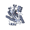
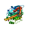
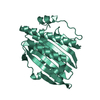
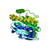
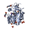
| Title | Crystal structure of PcyA V225D-biliverdin XIII alpha complex | ||||||
 Components Components | Phycocyanobilin:ferredoxin oxidoreductase | ||||||
 Keywords Keywords | OXIDOREDUCTASE / alpha/beta/alpha sandwich | ||||||
| Function / homology |  Function and homology information Function and homology informationphycocyanobilin:ferredoxin oxidoreductase / phycocyanobilin:ferredoxin oxidoreductase activity / phytochromobilin biosynthetic process / cobalt ion binding Similarity search - Function | ||||||
| Biological species |  | ||||||
| Method |  X-RAY DIFFRACTION / SYNCHROTRON / 3AJG / Resolution: 2.25 Å X-RAY DIFFRACTION / SYNCHROTRON / 3AJG / Resolution: 2.25 Å | ||||||
 Authors Authors | Wada, K. / Hagiwara, Y. / Fukuyama, K. | ||||||
 Citation Citation | Journal: Biochem.Biophys.Res.Commun. / Year: 2010 Title: One residue substitution in PcyA leads to unexpected changes in tetrapyrrole substrate binding. Authors: Wada, K. / Hagiwara, Y. / Yutani, Y. / Fukuyama, K. | ||||||
| History |
|
- Structure visualization
| Structure viewer | Molecule: MolmilJmol/JSmol |
|---|
- Downloads & links
-Download
| PDBx/mmCIF format | 3ajh.cif.gz | 112.2 KB | Display | PDBx/mmCIF format |
|---|---|---|---|---|
| PDB format | pdb3ajh.ent.gz | 88.3 KB | Display | PDB format |
| PDBx/mmJSON format | 3ajh.json.gz | Tree view | PDBx/mmJSON format | |
| Others |  Other downloads Other downloads |
-Validation report
| Arichive directory | https://data.pdbj.org/pub/pdb/validation_reports/aj/3ajhftp://data.pdbj.org/pub/pdb/validation_reports/aj/3ajh | HTTPS FTP |
|---|
-Related structure data


-Links
 PDBj
PDBj- Assembly
| Deposited unit | 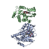
| ||||||||
|---|---|---|---|---|---|---|---|---|---|
| 1 | 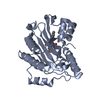
| ||||||||
| 2 | 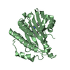
| ||||||||
| Unit cell |
|
-Components
| #1: Protein | Mass: 28172.113 Da / Num. of mol.: 2 / Fragment: V225D Source method: isolated from a genetically manipulated source Source: (gene. exp.) References: UniProt: Q55891, phycocyanobilin:ferredoxin oxidoreductase #2: Chemical |   Mass: 582.646 Da / Num. of mol.: 2 / Source method: obtained synthetically / Formula: C33H34N4O6 Mass: 582.646 Da / Num. of mol.: 2 / Source method: obtained synthetically / Formula: C33H34N4O6#3: Water | ChemComp-HOH / |  Mass: 18.015 Da / Num. of mol.: 134 / Source method: isolated from a natural source / Formula: H2O Mass: 18.015 Da / Num. of mol.: 134 / Source method: isolated from a natural source / Formula: H2O |
|---|
-Experimental details
-Experiment
| Experiment | Method: X-RAY DIFFRACTION / Number of used crystals: 1 |
|---|
- Sample preparation
| Crystal | Density Matthews: 2.09 Å3/Da / Density % sol: 41.15 % |
|---|---|
| Crystal grow | Temperature: 293 K / Method: vapor diffusion, hanging drop / pH: 4 Details: 0.8-1.0M NaH2PO4, 1.0-1.2M K2HPO4, 100mM sodium acetate, pH 4.0, VAPOR DIFFUSION, HANGING DROP, temperature 293K |
-Data collection
| Diffraction | Mean temperature: 100 K |
|---|---|
| Diffraction source | Source: SYNCHROTRON / Site: SPring-8  / Beamline: BL41XU / Wavelength: 1 Å / Beamline: BL41XU / Wavelength: 1 Å |
| Detector | Type: ADSC QUANTUM 315 / Detector: CCD / Date: Dec 11, 2009 |
| Radiation | Protocol: SINGLE WAVELENGTH / Monochromatic (M) / Laue (L): M / Scattering type: x-ray |
| Radiation wavelength | Wavelength: 1 Å / Relative weight: 1 |
| Reflection | Resolution: 2.25→50 Å / Num. all: 23018 / Num. obs: 23018 / % possible obs: 98.5 % / Observed criterion σ(F): 0 / Observed criterion σ(I): 0 / Redundancy: 7.4 % / Biso Wilson estimate: 29.2 Å2 / Rmerge(I) obs: 0.046 / Net I/σ(I): 18.5 |
| Reflection shell | Resolution: 2.25→2.29 Å / Redundancy: 7.4 % / Rmerge(I) obs: 0.31 / Num. unique all: 1118 / % possible all: 99.6 |
- Processing
| Software |
| |||||||||||||||||||||||||
|---|---|---|---|---|---|---|---|---|---|---|---|---|---|---|---|---|---|---|---|---|---|---|---|---|---|---|
| Refinement | Method to determine structure: 3AJG / Resolution: 2.25→27.98 Å / Rfactor Rfree error: 0.009 / Data cutoff high absF: 281864.72 / Data cutoff low absF: 0 / Isotropic thermal model: RESTRAINED / Cross valid method: THROUGHOUT / σ(F): 0 / Stereochemistry target values: Engh & Huber / Details: BULK SOLVENT MODEL USED
| |||||||||||||||||||||||||
| Solvent computation | Solvent model: FLAT MODEL / Bsol: 50.9269 Å2 / ksol: 0.35 e/Å3 | |||||||||||||||||||||||||
| Displacement parameters | Biso mean: 55.4 Å2
| |||||||||||||||||||||||||
| Refine analyze |
| |||||||||||||||||||||||||
| Refinement step | Cycle: LAST / Resolution: 2.25→27.98 Å
| |||||||||||||||||||||||||
| Refine LS restraints |
| |||||||||||||||||||||||||
| LS refinement shell | Resolution: 2.25→2.39 Å / Rfactor Rfree error: 0.031 / Total num. of bins used: 6
| |||||||||||||||||||||||||
| Xplor file |
|
