 Movie
Movie Controller

[English] 日本語
 Yorodumi
- PDB-6u9v: Cryo electron microscopy structure of the ATP-gated rat P2X7 ion ... -
+ Open data

- Basic information
| Entry | Database: PDB / ID: 6u9v | ||||||||||||
|---|---|---|---|---|---|---|---|---|---|---|---|---|---|
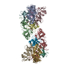
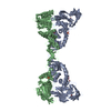
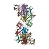
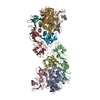
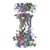
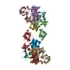
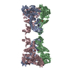
| Title | Cryo electron microscopy structure of the ATP-gated rat P2X7 ion channel in the apo, closed state | ||||||||||||
 Components Components | P2X purinoceptor 7 | ||||||||||||
 Keywords Keywords | MEMBRANE PROTEIN / Ion Channel Apoptosis | ||||||||||||
| Function / homology |  Function and homology information Function and homology informationPlatelet homeostasis / The NLRP3 inflammasome / positive regulation of lymphocyte apoptotic process / regulation of presynaptic dense core granule exocytosis / positive regulation of bleb assembly / NAD transport / Elevation of cytosolic Ca2+ levels / phagolysosome assembly / phospholipid transfer to membrane / positive regulation of cytoskeleton organization ...Platelet homeostasis / The NLRP3 inflammasome / positive regulation of lymphocyte apoptotic process / regulation of presynaptic dense core granule exocytosis / positive regulation of bleb assembly / NAD transport / Elevation of cytosolic Ca2+ levels / phagolysosome assembly / phospholipid transfer to membrane / positive regulation of cytoskeleton organization / positive regulation of monoatomic ion transmembrane transport / purinergic nucleotide receptor signaling pathway / plasma membrane organization / extracellularly ATP-gated monoatomic cation channel activity / positive regulation of interleukin-1 alpha production / purinergic nucleotide receptor activity / ATP export / collagen metabolic process / positive regulation of prostaglandin secretion / pore complex assembly / negative regulation of cell volume / plasma membrane phospholipid scrambling / positive regulation of gamma-aminobutyric acid secretion / bleb assembly / vesicle budding from membrane / positive regulation of T cell apoptotic process / bleb / response to fluid shear stress / programmed cell death / positive regulation of ossification / cell volume homeostasis / cellular response to dsRNA / negative regulation of bone resorption / ceramide biosynthetic process / positive regulation of macrophage cytokine production / skeletal system morphogenesis / phospholipid translocation / response to zinc ion / positive regulation of glutamate secretion / sodium channel activity / protein homotrimerization / response to ATP / T cell homeostasis / positive regulation of mitochondrial depolarization / membrane protein ectodomain proteolysis / positive regulation of NLRP3 inflammasome complex assembly / response to electrical stimulus / positive regulation of calcium ion transport into cytosol / synaptic vesicle exocytosis / T cell proliferation / positive regulation of bone mineralization / monoatomic cation transport / potassium channel activity / membrane depolarization / response to mechanical stimulus / regulation of sodium ion transport / neuronal action potential / extrinsic apoptotic signaling pathway / negative regulation of MAPK cascade / release of sequestered calcium ion into cytosol / reactive oxygen species metabolic process / homeostasis of number of cells within a tissue / sensory perception of pain / establishment of localization in cell / positive regulation of glycolytic process / positive regulation of interleukin-1 beta production / protein serine/threonine kinase activator activity / protein catabolic process / positive regulation of protein secretion / response to bacterium / neuromuscular junction / mitochondrion organization / apoptotic signaling pathway / lipopolysaccharide binding / protein processing / response to calcium ion / positive regulation of T cell mediated cytotoxicity / positive regulation of interleukin-6 production / calcium ion transmembrane transport / cell morphogenesis / cell-cell junction / terminal bouton / calcium ion transport / nuclear envelope / channel activity / signaling receptor activity / scaffold protein binding / response to lipopolysaccharide / gene expression / positive regulation of MAPK cascade / cell surface receptor signaling pathway / postsynapse / defense response to Gram-positive bacterium / positive regulation of apoptotic process / response to xenobiotic stimulus / inflammatory response / copper ion binding / signaling receptor binding / external side of plasma membrane / neuronal cell body Similarity search - Function | ||||||||||||
| Biological species |  | ||||||||||||
| Method | ELECTRON MICROSCOPY / single particle reconstruction / cryo EM / Resolution: 2.9 Å | ||||||||||||
 Authors Authors | Mansoor, S.E. / McCarthy, A.E. | ||||||||||||
| Funding support |  United States, 1items United States, 1items
| ||||||||||||
 Citation Citation | Journal: Cell / Year: 2019 Title: Full-Length P2X Structures Reveal How Palmitoylation Prevents Channel Desensitization. Authors: Alanna E McCarthy / Craig Yoshioka / Steven E Mansoor / Abstract: P2X receptors are trimeric, non-selective cation channels activated by extracellular ATP. The P2X receptor subtype is a pharmacological target because of involvement in apoptotic, inflammatory, and ...P2X receptors are trimeric, non-selective cation channels activated by extracellular ATP. The P2X receptor subtype is a pharmacological target because of involvement in apoptotic, inflammatory, and tumor progression pathways. It is the most structurally and functionally distinct P2X subtype, containing a unique cytoplasmic domain critical for the receptor to initiate apoptosis and not undergo desensitization. However, lack of structural information about the cytoplasmic domain has hindered understanding of the molecular mechanisms underlying these processes. We report cryoelectron microscopy structures of full-length rat P2X receptor in apo and ATP-bound states. These structures reveal how one cytoplasmic element, the C-cys anchor, prevents desensitization by anchoring the pore-lining helix to the membrane with palmitoyl groups. They show a second cytoplasmic element with a unique fold, the cytoplasmic ballast, which unexpectedly contains a zinc ion complex and a guanosine nucleotide binding site. Our structures provide first insights into the architecture and function of a P2X receptor cytoplasmic domain. | ||||||||||||
| History |
|
- Structure visualization
| Movie |
Movie viewer |
|---|---|
| Structure viewer | Molecule: MolmilJmol/JSmol |
- Downloads & links
-Download
| PDBx/mmCIF format | 6u9v.cif.gz | 580.2 KB | Display | PDBx/mmCIF format |
|---|---|---|---|---|
| PDB format | pdb6u9v.ent.gz | 489.3 KB | Display | PDB format |
| PDBx/mmJSON format | 6u9v.json.gz | Tree view | PDBx/mmJSON format | |
| Others |  Other downloads Other downloads |
-Validation report
| Arichive directory | https://data.pdbj.org/pub/pdb/validation_reports/u9/6u9vftp://data.pdbj.org/pub/pdb/validation_reports/u9/6u9v | HTTPS FTP |
|---|
-Related structure data
| Related structure data |  20702MC  6u9wC M: map data used to model this data C: citing same article ( |
|---|---|
| Similar structure data |

-Links
 PDBj
PDBj


- Assembly
| Deposited unit | 
|
|---|---|
| 1 |
|
-Components
-Protein , 1 types, 3 molecules ABC
| #1: Protein | Mass: 69904.016 Da / Num. of mol.: 3 Source method: isolated from a genetically manipulated source Source: (gene. exp.)  Homo sapiens (human) / References: UniProt: Q64663 Homo sapiens (human) / References: UniProt: Q64663 |
|---|
-Sugars , 3 types, 18 molecules 
| #2: Polysaccharide | 2-acetamido-2-deoxy-beta-D-glucopyranose-(1-4)-2-acetamido-2-deoxy-beta-D-glucopyranose Source method: isolated from a genetically manipulated source #3: Polysaccharide |   Source method: isolated from a genetically manipulated source Details: oligosaccharide / References: alpha-maltose #6: Sugar | ChemComp-NAG /  Type: D-saccharide, beta linking / Mass: 221.208 Da / Num. of mol.: 9 Type: D-saccharide, beta linking / Mass: 221.208 Da / Num. of mol.: 9Source method: isolated from a genetically manipulated source Formula: C8H15NO6 |
|---|
-Non-polymers , 4 types, 30 molecules 


| #4: Chemical |  Type: RNA linking / Mass: 443.201 Da / Num. of mol.: 3 / Source method: obtained synthetically / Formula: C10H15N5O11P2 / Feature type: SUBJECT OF INVESTIGATION / Comment: GDP, energy-carrying molecule*YM Type: RNA linking / Mass: 443.201 Da / Num. of mol.: 3 / Source method: obtained synthetically / Formula: C10H15N5O11P2 / Feature type: SUBJECT OF INVESTIGATION / Comment: GDP, energy-carrying molecule*YM#5: Chemical | ChemComp-ZN /  Mass: 65.409 Da / Num. of mol.: 6 / Source method: obtained synthetically / Formula: Zn / Feature type: SUBJECT OF INVESTIGATION Mass: 65.409 Da / Num. of mol.: 6 / Source method: obtained synthetically / Formula: Zn / Feature type: SUBJECT OF INVESTIGATION#7: Chemical |  Mass: 764.022 Da / Num. of mol.: 3 / Source method: obtained synthetically / Formula: C40H78NO10P / Comment: phospholipid*YM Mass: 764.022 Da / Num. of mol.: 3 / Source method: obtained synthetically / Formula: C40H78NO10P / Comment: phospholipid*YM#8: Chemical | ChemComp-PLM /  Mass: 256.424 Da / Num. of mol.: 18 / Source method: obtained synthetically / Formula: C16H32O2 Mass: 256.424 Da / Num. of mol.: 18 / Source method: obtained synthetically / Formula: C16H32O2 |
|---|
-Details
| Has ligand of interest | Y |
|---|---|
| Has protein modification | Y |
-Experimental details
-Experiment
| Experiment | Method: ELECTRON MICROSCOPY |
|---|---|
| EM experiment | Aggregation state: PARTICLE / 3D reconstruction method: single particle reconstruction |
- Sample preparation
| Component | Name: P2X7 receptor ion channel / Type: COMPLEX / Entity ID: #1 / Source: RECOMBINANT |
|---|---|
| Molecular weight | Experimental value: NO |
| Source (natural) | Organism: |
| Source (recombinant) | Organism: Homo sapiens (human) |
| Buffer solution | pH: 7 |
| Specimen | Embedding applied: NO / Shadowing applied: NO / Staining applied: NO / Vitrification applied: YES |
| Specimen support | Details: unspecified |
| Vitrification | Cryogen name: ETHANE |
- Electron microscopy imaging
| Experimental equipment |  Model: Titan Krios / Image courtesy: FEI Company |
|---|---|
| Microscopy | Model: FEI TITAN KRIOS |
| Electron gun | Electron source:  FIELD EMISSION GUN / Accelerating voltage: 300 kV / Illumination mode: FLOOD BEAM FIELD EMISSION GUN / Accelerating voltage: 300 kV / Illumination mode: FLOOD BEAM |
| Electron lens | Mode: BRIGHT FIELD |
| Image recording | Electron dose: 27 e/Å2 / Film or detector model: FEI FALCON III (4k x 4k) |
- Processing
| CTF correction | Type: PHASE FLIPPING ONLY |
|---|---|
| 3D reconstruction | Resolution: 2.9 Å / Resolution method: FSC 0.143 CUT-OFF / Num. of particles: 77697 / Symmetry type: POINT |
