Platelet homeostasis / The NLRP3 inflammasome / positive regulation of lymphocyte apoptotic process / regulation of presynaptic dense core granule exocytosis / positive regulation of bleb assembly / NAD transport / Elevation of cytosolic Ca2+ levels / phagolysosome assembly / phospholipid transfer to membrane / positive regulation of cytoskeleton organization ...Platelet homeostasis / The NLRP3 inflammasome / positive regulation of lymphocyte apoptotic process / regulation of presynaptic dense core granule exocytosis / positive regulation of bleb assembly / NAD transport / Elevation of cytosolic Ca2+ levels / phagolysosome assembly / phospholipid transfer to membrane / positive regulation of cytoskeleton organization / positive regulation of monoatomic ion transmembrane transport / purinergic nucleotide receptor signaling pathway / plasma membrane organization / extracellularly ATP-gated monoatomic cation channel activity / positive regulation of interleukin-1 alpha production / purinergic nucleotide receptor activity / collagen metabolic process / ATP export / positive regulation of prostaglandin secretion / pore complex assembly / negative regulation of cell volume / plasma membrane phospholipid scrambling / positive regulation of gamma-aminobutyric acid secretion / bleb assembly / vesicle budding from membrane / positive regulation of T cell apoptotic process / bleb / response to fluid shear stress / programmed cell death / positive regulation of ossification / cell volume homeostasis / cellular response to dsRNA / ceramide biosynthetic process / negative regulation of bone resorption / skeletal system morphogenesis / positive regulation of macrophage cytokine production / phospholipid translocation / response to zinc ion / positive regulation of glutamate secretion / protein homotrimerization / response to ATP / sodium channel activity / T cell homeostasis / membrane protein ectodomain proteolysis / positive regulation of mitochondrial depolarization / positive regulation of NLRP3 inflammasome complex assembly / positive regulation of calcium ion transport into cytosol / synaptic vesicle exocytosis / response to electrical stimulus / T cell proliferation / positive regulation of bone mineralization / monoatomic cation transport / potassium channel activity / membrane depolarization / response to mechanical stimulus / regulation of sodium ion transport / extrinsic apoptotic signaling pathway / neuronal action potential / negative regulation of MAPK cascade / release of sequestered calcium ion into cytosol / reactive oxygen species metabolic process / establishment of localization in cell / homeostasis of number of cells within a tissue / sensory perception of pain / positive regulation of glycolytic process / positive regulation of interleukin-1 beta production / protein catabolic process / protein serine/threonine kinase activator activity / positive regulation of protein secretion / response to bacterium / neuromuscular junction / mitochondrion organization / apoptotic signaling pathway / lipopolysaccharide binding / protein processing / response to calcium ion / positive regulation of T cell mediated cytotoxicity / positive regulation of interleukin-6 production / cell morphogenesis / calcium ion transmembrane transport / cell-cell junction / terminal bouton / calcium ion transport / nuclear envelope / channel activity / signaling receptor activity / scaffold protein binding / response to lipopolysaccharide / gene expression / positive regulation of MAPK cascade / cell surface receptor signaling pathway / postsynapse / defense response to Gram-positive bacterium / positive regulation of apoptotic process / response to xenobiotic stimulus / inflammatory response / copper ion binding / signaling receptor binding / external side of plasma membrane / neuronal cell body Similarity search - Function
National Institutes of Health/National Heart, Lung, and Blood Institute (NIH/NHLBI)
K99HL138129
United States
Citation
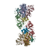
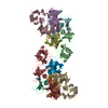
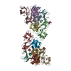
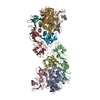
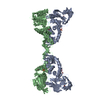
Journal: Cell / Year: 2019 Title: Full-Length P2X Structures Reveal How Palmitoylation Prevents Channel Desensitization. Authors: Alanna E McCarthy / Craig Yoshioka / Steven E Mansoor / Abstract: P2X receptors are trimeric, non-selective cation channels activated by extracellular ATP. The P2X receptor subtype is a pharmacological target because of involvement in apoptotic, inflammatory, and ...P2X receptors are trimeric, non-selective cation channels activated by extracellular ATP. The P2X receptor subtype is a pharmacological target because of involvement in apoptotic, inflammatory, and tumor progression pathways. It is the most structurally and functionally distinct P2X subtype, containing a unique cytoplasmic domain critical for the receptor to initiate apoptosis and not undergo desensitization. However, lack of structural information about the cytoplasmic domain has hindered understanding of the molecular mechanisms underlying these processes. We report cryoelectron microscopy structures of full-length rat P2X receptor in apo and ATP-bound states. These structures reveal how one cytoplasmic element, the C-cys anchor, prevents desensitization by anchoring the pore-lining helix to the membrane with palmitoyl groups. They show a second cytoplasmic element with a unique fold, the cytoplasmic ballast, which unexpectedly contains a zinc ion complex and a guanosine nucleotide binding site. Our structures provide first insights into the architecture and function of a P2X receptor cytoplasmic domain.
History
Deposition
Sep 9, 2019
-
Header (metadata) release
Oct 2, 2019
-
Map release
Oct 23, 2019
-
Update
Nov 20, 2024
-
Current status
Nov 20, 2024
Processing site: RCSB / Status: Released
-
Structure visualization
Movie
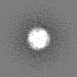
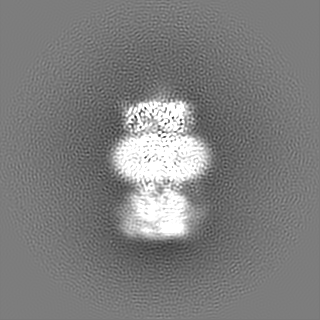
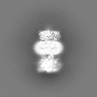
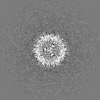
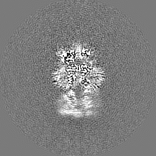
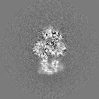
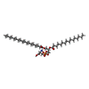
Surface view with section colored by density value
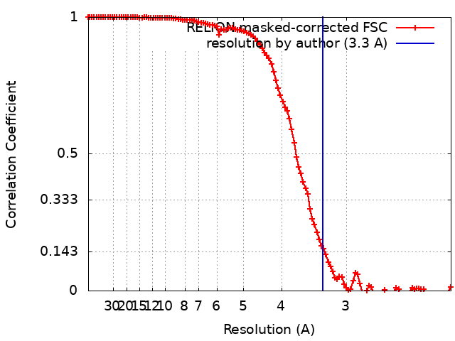
Map for the ATP-bound (open) state of rat P2X7 receptor from a focused refinement using a mask that includes the transmembrane domain and the cytoplasmic domain but excludes the extracellular domain.
In the structure databanks used in Yorodumi, some data are registered as the other names, "COVID-19 virus" and "2019-nCoV". Here are the details of the virus and the list of structure data.
Jan 31, 2019. EMDB accession codes are about to change! (news from PDBe EMDB page)
EMDB accession codes are about to change! (news from PDBe EMDB page)
The allocation of 4 digits for EMDB accession codes will soon come to an end. Whilst these codes will remain in use, new EMDB accession codes will include an additional digit and will expand incrementally as the available range of codes is exhausted. The current 4-digit format prefixed with “EMD-” (i.e. EMD-XXXX) will advance to a 5-digit format (i.e. EMD-XXXXX), and so on. It is currently estimated that the 4-digit codes will be depleted around Spring 2019, at which point the 5-digit format will come into force.
The EM Navigator/Yorodumi systems omit the EMD- prefix.
Related info.:Q: What is EMD? / ID/Accession-code notation in Yorodumi/EM Navigator
Yorodumi is a browser for structure data from EMDB, PDB, SASBDB, etc.
This page is also the successor to EM Navigator detail page, and also detail information page/front-end page for Omokage search.
The word "yorodu" (or yorozu) is an old Japanese word meaning "ten thousand". "mi" (miru) is to see.
Related info.:EMDB / PDB / SASBDB / Comparison of 3 databanks / Yorodumi Search / Aug 31, 2016. New EM Navigator & Yorodumi / Yorodumi Papers / Jmol/JSmol / Function and homology information / Changes in new EM Navigator and Yorodumi
 Movie
Movie Controller

 Yorodumi
 Open data
 Basic information
 Map data
 Sample
 Keywords
 Function and homology information
 Authors
 United States, 1 items
  Citation
 Structure visualization
 Downloads & links
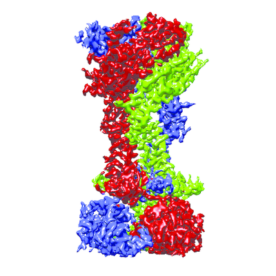 emd_20703.png
 http://ftp.pdbj.org/pub/emdb/structures/EMD-20703


 Z (Sec.)
 Y (Row.)
 X (Col.)


 Sample components
 Homo sapiens (human)


 Processing
 Electron microscopy
 FIELD EMISSION GUN