 Movie
Movie Controller

[English] 日本語
 Yorodumi
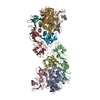
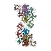
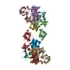
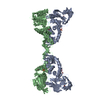
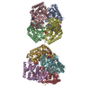
- PDB-6ro0: CRYSTAL STRUCTURE OF GENETICALLY DETOXIFIED PERTUSSIS TOXIN GDPT. -
+ Open data

- Basic information
| Entry | Database: PDB / ID: 6ro0 | ||||||
|---|---|---|---|---|---|---|---|
| Title | CRYSTAL STRUCTURE OF GENETICALLY DETOXIFIED PERTUSSIS TOXIN GDPT. | ||||||
 Components Components |
| ||||||
 Keywords Keywords | TOXIN | ||||||
| Function / homology |  Function and homology information Function and homology informationsymbiont-mediated activation of host MAPK cascade / symbiont-mediated activation of host G protein-coupled receptor signal transduction / Transferases; Glycosyltransferases; Pentosyltransferases / NAD+ poly-ADP-ribosyltransferase activity / nucleotidyltransferase activity / toxin activity / host cell plasma membrane / extracellular region / plasma membrane Similarity search - Function | ||||||
| Biological species |  Bordetella pertussis (bacteria) Bordetella pertussis (bacteria) | ||||||
| Method |  X-RAY DIFFRACTION / SYNCHROTRON / MOLECULAR REPLACEMENT / Resolution: 2.13 Å X-RAY DIFFRACTION / SYNCHROTRON / MOLECULAR REPLACEMENT / Resolution: 2.13 Å | ||||||
 Authors Authors | Bertrand, T. | ||||||
 Citation Citation | Journal: Commun Biol / Year: 2020 Title: Genetically detoxified pertussis toxin displays near identical structure to its wild-type and exhibits robust immunogenicity. Authors: Ausar, S.F. / Zhu, S. / Duprez, J. / Cohen, M. / Bertrand, T. / Steier, V. / Wilson, D.J. / Li, S. / Sheung, A. / Brookes, R.H. / Pedyczak, A. / Rak, A. / Andrew James, D. | ||||||
| History |
|
- Structure visualization
| Structure viewer | Molecule: MolmilJmol/JSmol |
|---|
- Downloads & links
-Download
| PDBx/mmCIF format | 6ro0.cif.gz | 393.9 KB | Display | PDBx/mmCIF format |
|---|---|---|---|---|
| PDB format | pdb6ro0.ent.gz | 311.8 KB | Display | PDB format |
| PDBx/mmJSON format | 6ro0.json.gz | Tree view | PDBx/mmJSON format | |
| Others |  Other downloads Other downloads |
-Validation report
| Arichive directory | https://data.pdbj.org/pub/pdb/validation_reports/ro/6ro0ftp://data.pdbj.org/pub/pdb/validation_reports/ro/6ro0 | HTTPS FTP |
|---|
-Related structure data
| Related structure data |  1prtS S: Starting model for refinement |
|---|---|
| Similar structure data |


-Links
 PDBj
PDBj


- Assembly
| Deposited unit | 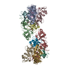
| ||||||||
|---|---|---|---|---|---|---|---|---|---|
| 1 |
| ||||||||
| Unit cell |
|
-Components
-Pertussis toxin subunit ... , 2 types, 4 molecules AGFL
| #1: Protein | Mass: 29905.109 Da / Num. of mol.: 2 / Mutation: R9K, E129G Source method: isolated from a genetically manipulated source Source: (gene. exp.) Bordetella pertussis (bacteria) / Gene: ptxA / Production host: Bordetella pertussis (bacteria) / References: UniProt: T1SR96, UniProt: P04977*PLUS#5: Protein | Mass: 14513.755 Da / Num. of mol.: 2 Source method: isolated from a genetically manipulated source Source: (gene. exp.) Bordetella pertussis (bacteria)Gene: ptxE, ERS004697_00105, ERS004704_00093, ERS004709_00175, ERS005128_00197, ERS005179_00180, ERS014547_00748, ERS014550_01624, ERS018341_00090 Production host: Bordetella pertussis (bacteria) / References: UniProt: C0MPK9, UniProt: P04981*PLUS |
|---|
-Islet-activating protein ... , 3 types, 8 molecules BHCIDEJK
| #2: Protein | Mass: 24856.299 Da / Num. of mol.: 2 Source method: isolated from a genetically manipulated source Source: (gene. exp.) Bordetella pertussis (bacteria)Gene: ptxB, CYO81_20230, ERS004697_00332, ERS004704_00067, ERS004709_00223, ERS005128_00199, ERS005179_00182, ERS014547_00750, NCTC10911_00247 Production host: Bordetella pertussis (bacteria) / References: UniProt: A0A0E8DFW5, UniProt: P04978*PLUS#3: Protein | Mass: 25012.820 Da / Num. of mol.: 2 Source method: isolated from a genetically manipulated source Source: (gene. exp.) Bordetella pertussis (bacteria)Gene: ptxS3, ptxC, ptxC_1, ptxC_2, CYO81_20245, ERS004697_00106, ERS004704_00094, ERS004709_00174, ERS005128_00196, ERS005179_00179, ERS014547_00747, ERS014550_01623, NCTC10911_00250 Production host: Bordetella pertussis (bacteria) / References: UniProt: Q546I1, UniProt: P04979*PLUS#4: Protein | Mass: 16566.744 Da / Num. of mol.: 4 Source method: isolated from a genetically manipulated source Source: (gene. exp.) Bordetella pertussis (bacteria)Gene: ptxD, CYO81_20235, ERS005128_00198, ERS005179_00181, ERS014547_00749, NCTC10911_00248 Production host: Bordetella pertussis (bacteria) / References: UniProt: C0MPK8, UniProt: P0A3R5*PLUS |
|---|
-Non-polymers , 2 types, 533 molecules 

| #6: Chemical | ChemComp-GOL /  Mass: 92.094 Da / Num. of mol.: 9 / Source method: obtained synthetically / Formula: C3H8O3 Mass: 92.094 Da / Num. of mol.: 9 / Source method: obtained synthetically / Formula: C3H8O3#7: Water | ChemComp-HOH / | Mass: 18.015 Da / Num. of mol.: 524 / Source method: isolated from a natural source / Formula: H2O |
|---|
-Details
| Has protein modification | Y |
|---|
-Experimental details
-Experiment
| Experiment | Method: X-RAY DIFFRACTION / Number of used crystals: 1 |
|---|
- Sample preparation
| Crystal | Density Matthews: 2.9 Å3/Da / Density % sol: 57.63 % |
|---|---|
| Crystal grow | Temperature: 278 K / Method: evaporation / pH: 7 / Details: Spontaneous crystallization in plastic bottles / PH range: 7 |
-Data collection
| Diffraction | Mean temperature: 100 K / Serial crystal experiment: N |
|---|---|
| Diffraction source | Source: SYNCHROTRON / Site: ESRF  / Beamline: MASSIF-1 / Wavelength: 0.966 Å / Beamline: MASSIF-1 / Wavelength: 0.966 Å |
| Detector | Type: DECTRIS PILATUS3 2M / Detector: PIXEL / Date: Apr 26, 2017 |
| Radiation | Protocol: SINGLE WAVELENGTH / Monochromatic (M) / Laue (L): M / Scattering type: x-ray |
| Radiation wavelength | Wavelength: 0.966 Å / Relative weight: 1 |
| Reflection | Resolution: 2.13→46.96 Å / Num. obs: 165565 / % possible obs: 99.8 % / Redundancy: 4.8 % / Biso Wilson estimate: 50.68 Å2 / Rmerge(I) obs: 0.065 / Net I/σ(I): 11.5 |
| Reflection shell | Resolution: 2.13→2.2 Å / Rmerge(I) obs: 1.95 / Num. unique obs: 16119 |
- Processing
| Software |
| ||||||||||||||||||||||||||||||||||||||||||||||||||||||||||||||||||||||||||||||||||||||||||||||||||||||||||||||||||
|---|---|---|---|---|---|---|---|---|---|---|---|---|---|---|---|---|---|---|---|---|---|---|---|---|---|---|---|---|---|---|---|---|---|---|---|---|---|---|---|---|---|---|---|---|---|---|---|---|---|---|---|---|---|---|---|---|---|---|---|---|---|---|---|---|---|---|---|---|---|---|---|---|---|---|---|---|---|---|---|---|---|---|---|---|---|---|---|---|---|---|---|---|---|---|---|---|---|---|---|---|---|---|---|---|---|---|---|---|---|---|---|---|---|---|---|
| Refinement | Method to determine structure: MOLECULAR REPLACEMENT Starting model: 1PRT Resolution: 2.13→46.62 Å / Cor.coef. Fo:Fc: 0.904 / Cor.coef. Fo:Fc free: 0.89 / SU R Cruickshank DPI: 0.194 / Cross valid method: THROUGHOUT / σ(F): 0 / SU R Blow DPI: 0.192 / SU Rfree Blow DPI: 0.165 / SU Rfree Cruickshank DPI: 0.167
| ||||||||||||||||||||||||||||||||||||||||||||||||||||||||||||||||||||||||||||||||||||||||||||||||||||||||||||||||||
| Displacement parameters | Biso mean: 64.94 Å2
| ||||||||||||||||||||||||||||||||||||||||||||||||||||||||||||||||||||||||||||||||||||||||||||||||||||||||||||||||||
| Refine analyze | Luzzati coordinate error obs: 0.44 Å | ||||||||||||||||||||||||||||||||||||||||||||||||||||||||||||||||||||||||||||||||||||||||||||||||||||||||||||||||||
| Refinement step | Cycle: LAST / Resolution: 2.13→46.62 Å
| ||||||||||||||||||||||||||||||||||||||||||||||||||||||||||||||||||||||||||||||||||||||||||||||||||||||||||||||||||
| Refine LS restraints |
| ||||||||||||||||||||||||||||||||||||||||||||||||||||||||||||||||||||||||||||||||||||||||||||||||||||||||||||||||||
| LS refinement shell | Resolution: 2.13→2.19 Å / Total num. of bins used: 20
|
