 Movie
Movie Controller


+ Open data

- Basic information
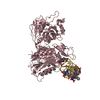
| Entry | Database: PDB / ID: 1bcp | ||||||
|---|---|---|---|---|---|---|---|
| Title | BINARY COMPLEX OF PERTUSSIS TOXIN AND ATP | ||||||
 Components Components | (PERTUSSIS TOXIN) x 5 | ||||||
 Keywords Keywords | TOXIN / ADP-RIBOSYLTRANSFERASE / TRANSFERASE / WHOOPING COUGH | ||||||
| Function / homology |  Function and homology information Function and homology informationsymbiont-mediated activation of host MAPK cascade / symbiont-mediated activation of host G protein-coupled receptor signal transduction / Transferases; Glycosyltransferases; Pentosyltransferases / NAD+ poly-ADP-ribosyltransferase activity / nucleotidyltransferase activity / toxin activity / host cell plasma membrane / extracellular region Similarity search - Function | ||||||
| Biological species |  Bordetella pertussis (bacteria) Bordetella pertussis (bacteria) | ||||||
| Method |  X-RAY DIFFRACTION / SYNCHROTRON / Resolution: 2.7 Å X-RAY DIFFRACTION / SYNCHROTRON / Resolution: 2.7 Å | ||||||
 Authors Authors | Hazes, B. / Read, R.J. | ||||||
 Citation Citation | Journal: J.Mol.Biol. / Year: 1996 Title: Crystal structure of the pertussis toxin-ATP complex: a molecular sensor. Authors: Hazes, B. / Boodhoo, A. / Cockle, S.A. / Read, R.J. #1: Journal: Structure / Year: 1994Title: The Crystal Structure of Pertussis Toxin Authors: Stein, P.E. / Boodhoo, A. / Armstrong, G.D. / Cockle, S.A. / Klein, M.H. / Read, R.J. #2: Journal: Nat.Struct.Biol. / Year: 1994Title: Structure of a Pertussis Toxin-Sugar Complex as a Model for Receptor Binding Authors: Stein, P.E. / Boodhoo, A. / Armstrong, G.D. / Heerze, L.D. / Cockle, S.A. / Klein, M.H. / Read, R.J. #3: Journal: Nucleic Acids Res. / Year: 1989Title: A Unique Sequence of the Bordetella Pertussis Toxin Operon Authors: Loosmore, S.M. / Cunningham, J.D. / Bradley, W.R. / Yao, F.L. / Dekaban, G.A. / Klein, M.H. | ||||||
| History |
|
- Structure visualization
| Structure viewer | Molecule: MolmilJmol/JSmol |
|---|
- Downloads & links
-Download
| PDBx/mmCIF format | 1bcp.cif.gz | 364.3 KB | Display | PDBx/mmCIF format |
|---|---|---|---|---|
| PDB format | pdb1bcp.ent.gz | 294.3 KB | Display | PDB format |
| PDBx/mmJSON format | 1bcp.json.gz | Tree view | PDBx/mmJSON format | |
| Others |  Other downloads Other downloads |
-Validation report
| Arichive directory | https://data.pdbj.org/pub/pdb/validation_reports/bc/1bcpftp://data.pdbj.org/pub/pdb/validation_reports/bc/1bcp | HTTPS FTP |
|---|
-Related structure data
| Similar structure data |
|---|


-Links
 PDBj
PDBj


- Assembly
| Deposited unit | 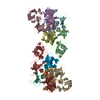
| ||||||||
|---|---|---|---|---|---|---|---|---|---|
| 1 | 
| ||||||||
| 2 | 
| ||||||||
| Unit cell |
| ||||||||
| Noncrystallographic symmetry (NCS) | NCS oper: (Code: given Matrix: (-0.91421, 0.372578, -0.159394), Vector: Details | EACH OF THE TWO HOLOTOXIN MOLECULES IN THE ASYMMETRIC UNIT CONSISTS OF SIX SUBUNITS AND THEY HAVE BEEN ASSIGNED CHAIN INDICATORS A - F AND G - L, RESPECTIVELY. THE TRANSFORMATION PRESENTED ON *MTRIX* RECORDS BELOW WILL YIELD APPROXIMATE COORDINATES FOR CHAINS G - L WHEN APPLIED TO CHAINS A - F. | |
-Components
-Protein , 5 types, 12 molecules AGBHCIDEJKFL
| #1: Protein | Mass: 26248.740 Da / Num. of mol.: 2 / Source method: isolated from a natural source Details: THE PERTUSSIS TOXIN USED FOR THIS WORK WAS PURIFIED FROM B. PERTUSSIS STRAIN 10536 (LOOSMORE ET AL., NUCLEIC ACIDS RES., VOL. 17, 8365, 1989), WHICH DIFFERS AT TWO POSITIONS IN SUBUNIT S1 ...Details: THE PERTUSSIS TOXIN USED FOR THIS WORK WAS PURIFIED FROM B. PERTUSSIS STRAIN 10536 (LOOSMORE ET AL., NUCLEIC ACIDS RES., VOL. 17, 8365, 1989), WHICH DIFFERS AT TWO POSITIONS IN SUBUNIT S1 (ASP 34 GLU AND ILE 198 VAL) FROM THE SEQUENCE THAT WAS FIRST REPORTED FOR THE PROTEIN (NICOSIA ET AL., PNAS VOL 83, 4631 - 4635, 1986). Source: (natural) Bordetella pertussis (bacteria) / Strain: 10536References: UniProt: P04977, Transferases; Glycosyltransferases; Pentosyltransferases #2: Protein | Mass: 21943.701 Da / Num. of mol.: 2 / Source method: isolated from a natural source Details: THE PERTUSSIS TOXIN USED FOR THIS WORK WAS PURIFIED FROM B. PERTUSSIS STRAIN 10536 (LOOSMORE ET AL., NUCLEIC ACIDS RES., VOL. 17, 8365, 1989), WHICH DIFFERS AT TWO POSITIONS IN SUBUNIT S1 ...Details: THE PERTUSSIS TOXIN USED FOR THIS WORK WAS PURIFIED FROM B. PERTUSSIS STRAIN 10536 (LOOSMORE ET AL., NUCLEIC ACIDS RES., VOL. 17, 8365, 1989), WHICH DIFFERS AT TWO POSITIONS IN SUBUNIT S1 (ASP 34 GLU AND ILE 198 VAL) FROM THE SEQUENCE THAT WAS FIRST REPORTED FOR THE PROTEIN (NICOSIA ET AL., PNAS VOL 83, 4631 - 4635, 1986). Source: (natural) Bordetella pertussis (bacteria) / Strain: 10536References: UniProt: P04978, Transferases; Glycosyltransferases; Pentosyltransferases #3: Protein | Mass: 21889.867 Da / Num. of mol.: 2 / Source method: isolated from a natural source Details: THE PERTUSSIS TOXIN USED FOR THIS WORK WAS PURIFIED FROM B. PERTUSSIS STRAIN 10536 (LOOSMORE ET AL., NUCLEIC ACIDS RES., VOL. 17, 8365, 1989), WHICH DIFFERS AT TWO POSITIONS IN SUBUNIT S1 ...Details: THE PERTUSSIS TOXIN USED FOR THIS WORK WAS PURIFIED FROM B. PERTUSSIS STRAIN 10536 (LOOSMORE ET AL., NUCLEIC ACIDS RES., VOL. 17, 8365, 1989), WHICH DIFFERS AT TWO POSITIONS IN SUBUNIT S1 (ASP 34 GLU AND ILE 198 VAL) FROM THE SEQUENCE THAT WAS FIRST REPORTED FOR THE PROTEIN (NICOSIA ET AL., PNAS VOL 83, 4631 - 4635, 1986). Source: (natural) Bordetella pertussis (bacteria) / Strain: 10536References: UniProt: P04979, Transferases; Glycosyltransferases; Pentosyltransferases #4: Protein | Mass: 12072.426 Da / Num. of mol.: 4 / Source method: isolated from a natural source Details: THE PERTUSSIS TOXIN USED FOR THIS WORK WAS PURIFIED FROM B. PERTUSSIS STRAIN 10536 (LOOSMORE ET AL., NUCLEIC ACIDS RES., VOL. 17, 8365, 1989), WHICH DIFFERS AT TWO POSITIONS IN SUBUNIT S1 ...Details: THE PERTUSSIS TOXIN USED FOR THIS WORK WAS PURIFIED FROM B. PERTUSSIS STRAIN 10536 (LOOSMORE ET AL., NUCLEIC ACIDS RES., VOL. 17, 8365, 1989), WHICH DIFFERS AT TWO POSITIONS IN SUBUNIT S1 (ASP 34 GLU AND ILE 198 VAL) FROM THE SEQUENCE THAT WAS FIRST REPORTED FOR THE PROTEIN (NICOSIA ET AL., PNAS VOL 83, 4631 - 4635, 1986). Source: (natural) Bordetella pertussis (bacteria) / Strain: 10536References: UniProt: P0A3R5, Transferases; Glycosyltransferases; Pentosyltransferases #5: Protein | Mass: 10951.524 Da / Num. of mol.: 2 / Source method: isolated from a natural source Details: THE PERTUSSIS TOXIN USED FOR THIS WORK WAS PURIFIED FROM B. PERTUSSIS STRAIN 10536 (LOOSMORE ET AL., NUCLEIC ACIDS RES., VOL. 17, 8365, 1989), WHICH DIFFERS AT TWO POSITIONS IN SUBUNIT S1 ...Details: THE PERTUSSIS TOXIN USED FOR THIS WORK WAS PURIFIED FROM B. PERTUSSIS STRAIN 10536 (LOOSMORE ET AL., NUCLEIC ACIDS RES., VOL. 17, 8365, 1989), WHICH DIFFERS AT TWO POSITIONS IN SUBUNIT S1 (ASP 34 GLU AND ILE 198 VAL) FROM THE SEQUENCE THAT WAS FIRST REPORTED FOR THE PROTEIN (NICOSIA ET AL., PNAS VOL 83, 4631 - 4635, 1986). Source: (natural) Bordetella pertussis (bacteria) / Strain: 10536References: UniProt: P04981, Transferases; Glycosyltransferases; Pentosyltransferases |
|---|
-Non-polymers , 2 types, 43 molecules 

| #6: Chemical |  Mass: 507.181 Da / Num. of mol.: 2 / Source method: obtained synthetically / Formula: C10H16N5O13P3 / Comment: ATP, energy-carrying molecule*YM Mass: 507.181 Da / Num. of mol.: 2 / Source method: obtained synthetically / Formula: C10H16N5O13P3 / Comment: ATP, energy-carrying molecule*YM#7: Water | ChemComp-HOH / | Mass: 18.015 Da / Num. of mol.: 41 / Source method: isolated from a natural source / Formula: H2O |
|---|
-Details
| Compound details | SUBUNIT S1 OF THE HOLOTOXIN MOLECULE (CHAINS A AND G) FORMS THE ENZYMATIC PART OF THE TOXIN. S1 ADP- ...SUBUNIT S1 OF THE HOLOTOXIN MOLECULE (CHAINS A AND G) FORMS THE ENZYMATIC PART OF THE TOXIN. S1 ADP-RIBOSYLATE |
|---|---|
| Has protein modification | Y |
| Nonpolymer details | ATP 1 IS BOUND TO HOLOTOXIN MOLECULE 1 (CHAINS A-F). ATP 2 IS BOUND TO HOLOTOXIN MOLECULE 2 (CHAINS ...ATP 1 IS BOUND TO HOLOTOXIN MOLECULE 1 (CHAINS A-F). ATP 2 IS BOUND TO HOLOTOXIN MOLECULE 2 (CHAINS G-L). WATER MOLECULES 3 TO 27 BIND TO HOLOTOXIN MOLECULE 1. WATER MOLECULES 28 TO 43 BIND TO HOLOTOXIN MOLECULE 2. |
| Sequence details | THE PERTUSSIS TOXIN USED FOR THIS WORK WAS PURIFIED FROM B. PERTUSSIS STRAIN 10536 (LOOSMORE ET AL. ...THE PERTUSSIS TOXIN USED FOR THIS WORK WAS PURIFIED FROM B. PERTUSSIS STRAIN 10536 (LOOSMORE ET AL., NUCLEIC ACIDS RES., VOL. 17, 8365, 1989), WHICH DIFFERS AT TWO POSITIONS IN SUBUNIT S1 (ASP 34 GLU AND ILE 198 VAL) FROM THE SEQUENCE THAT WAS FIRST REPORTED FOR THE PROTEIN (NICOSIA ET AL., PNAS VOL 83, 4631 - 4635, 1986). |
-Experimental details
-Experiment
| Experiment | Method: X-RAY DIFFRACTION |
|---|
- Sample preparation
| Crystal | Density Matthews: 3.72 Å3/Da / Density % sol: 66.9 % Description: DATA COLLECTION STATISTICS ARE GIVEN FOR ALL DATA UP TO 2.5 ANGSTROMS. HOWEVER, DUE TO RADIATION DAMAGE THE HIGH RESOLUTION DATA IS VERY INCOMPLETE AND THEREFORE ONLY DATA TO 2.7 ...Description: DATA COLLECTION STATISTICS ARE GIVEN FOR ALL DATA UP TO 2.5 ANGSTROMS. HOWEVER, DUE TO RADIATION DAMAGE THE HIGH RESOLUTION DATA IS VERY INCOMPLETE AND THEREFORE ONLY DATA TO 2.7 ANGSTROM HAVE BEEN USED FOR REFINEMENT. THE DATA UP TO THIS RESOLUTION IS 67.3 % COMPLETE WITH A COMPLETENESS OF 19.6 % IN THE HIGHEST RESOLUTION SHELL | ||||||||||||||||||||||||||||||
|---|---|---|---|---|---|---|---|---|---|---|---|---|---|---|---|---|---|---|---|---|---|---|---|---|---|---|---|---|---|---|---|
| Crystal grow | pH: 8 / Details: pH 8.0 | ||||||||||||||||||||||||||||||
| Crystal grow | *PLUS Method: vapor diffusion, hanging drop / Details: Stein, P.E., (1994) Structure (London), 2, 45. | ||||||||||||||||||||||||||||||
| Components of the solutions | *PLUS
|
-Data collection
| Diffraction | Mean temperature: 293 K |
|---|---|
| Diffraction source | Source: SYNCHROTRON / Site: Photon Factory  / Beamline: BL-6A / Wavelength: 1 / Beamline: BL-6A / Wavelength: 1 |
| Detector | Type: WEISSENBERG / Detector: DIFFRACTOMETER / Date: Nov 27, 1993 |
| Radiation | Monochromatic (M) / Laue (L): M / Scattering type: x-ray |
| Radiation wavelength | Wavelength: 1 Å / Relative weight: 1 |
| Reflection | Highest resolution: 2.5 Å / Num. obs: 62637 / % possible obs: 64 % / Observed criterion σ(I): 0 / Redundancy: 3 % / Rmerge(I) obs: 0.093 |
| Reflection | *PLUS Num. measured all: 195544 |
| Reflection shell | *PLUS Highest resolution: 2.5 Å / Lowest resolution: 2.7 Å / % possible obs: 19.6 % |
- Processing
| Software |
| ||||||||||||||||||||||||||||||||||||||||||||||||||||||||||||
|---|---|---|---|---|---|---|---|---|---|---|---|---|---|---|---|---|---|---|---|---|---|---|---|---|---|---|---|---|---|---|---|---|---|---|---|---|---|---|---|---|---|---|---|---|---|---|---|---|---|---|---|---|---|---|---|---|---|---|---|---|---|
| Refinement | Resolution: 2.7→8 Å / σ(F): 0 Details: THERE ARE MISSING RESIDUES AT THE N-TERMINI OF SUBUNITS S1, S2, S3, AND S5. IN ADDITION, NO COORDINATES ARE PRESENT FOR RESIDUES 211 - 220 IN SUBUNIT S1 (CHAINS A AND G). DATA COLLECTION ...Details: THERE ARE MISSING RESIDUES AT THE N-TERMINI OF SUBUNITS S1, S2, S3, AND S5. IN ADDITION, NO COORDINATES ARE PRESENT FOR RESIDUES 211 - 220 IN SUBUNIT S1 (CHAINS A AND G). DATA COLLECTION STATISTICS ARE GIVEN FOR ALL DATA UP TO 2.5 ANGSTROMS. HOWEVER, DUE TO RADIATION DAMAGE THE HIGH RESOLUTION DATA IS VERY INCOMPLETE AND THEREFORE ONLY DATA TO 2.7 ANGSTROM HAVE BEEN USED FOR REFINEMENT. THE DATA UP TO THIS RESOLUTION IS 67.3 % COMPLETE WITH A COMPLETENESS OF 19.6 % IN THE HIGHEST RESOLUTION SHELL
| ||||||||||||||||||||||||||||||||||||||||||||||||||||||||||||
| Displacement parameters | Biso mean: 21.84 Å2 | ||||||||||||||||||||||||||||||||||||||||||||||||||||||||||||
| Refine analyze | Luzzati sigma a obs: 0.46 Å | ||||||||||||||||||||||||||||||||||||||||||||||||||||||||||||
| Refinement step | Cycle: LAST / Resolution: 2.7→8 Å
| ||||||||||||||||||||||||||||||||||||||||||||||||||||||||||||
| Refine LS restraints |
|
