 Movie
Movie Controller

[English] 日本語
 Yorodumi
- PDB-1pto: THE STRUCTURE OF A PERTUSSIS TOXIN-SUGAR COMPLEX AS A MODEL FOR R... -
+ Open data

- Basic information
| Entry | Database: PDB / ID: 1pto | |||||||||
|---|---|---|---|---|---|---|---|---|---|---|
| Title | THE STRUCTURE OF A PERTUSSIS TOXIN-SUGAR COMPLEX AS A MODEL FOR RECEPTOR BINDING | |||||||||
 Components Components |
| |||||||||
 Keywords Keywords | TOXIN | |||||||||
| Function / homology |  Function and homology information Function and homology informationsymbiont-mediated activation of host MAPK cascade / symbiont-mediated activation of host G protein-coupled receptor signal transduction / : / Transferases; Glycosyltransferases; Pentosyltransferases / NAD+ poly-ADP-ribosyltransferase activity / nucleotidyltransferase activity / toxin activity / host cell plasma membrane / extracellular region / membrane Similarity search - Function | |||||||||
| Biological species |  Bordetella pertussis (bacteria) Bordetella pertussis (bacteria) | |||||||||
| Method |  X-RAY DIFFRACTION / Resolution: 3.5 Å X-RAY DIFFRACTION / Resolution: 3.5 Å | |||||||||
 Authors Authors | Stein, P.E. / Read, R.J. | |||||||||
 Citation Citation | Journal: Nat.Struct.Biol. / Year: 1994 Title: Structure of a pertussis toxin-sugar complex as a model for receptor binding. Authors: Stein, P.E. / Boodhoo, A. / Armstrong, G.D. / Heerze, L.D. / Cockle, S.A. / Klein, M.H. / Read, R.J. #1: Journal: Structure / Year: 1994Title: The Crystal Structure of Pertussis Toxin Authors: Stein, P.E. / Boodhoo, A. / Armstrong, G.D. / Cockle, S.A. / Klein, M.H. / Read, R.J. | |||||||||
| History |
| |||||||||
| Remark 700 | SHEET STRAND 2 MAKES HYDROGEN BONDS WITH STRAND 1 OF SHEET B5 AND ALSO WITH STRAND 2 OF SHEET B7. ...SHEET STRAND 2 MAKES HYDROGEN BONDS WITH STRAND 1 OF SHEET B5 AND ALSO WITH STRAND 2 OF SHEET B7. STRAND 2 MAKES HYDROGEN BONDS WITH STRAND 1 OF SHEET B9 AND ALSO WITH STRAND 2 OF SHEET B11. |
- Structure visualization
| Structure viewer | Molecule: MolmilJmol/JSmol |
|---|
- Downloads & links
-Download
| PDBx/mmCIF format | 1pto.cif.gz | 368.4 KB | Display | PDBx/mmCIF format |
|---|---|---|---|---|
| PDB format | pdb1pto.ent.gz | 296.3 KB | Display | PDB format |
| PDBx/mmJSON format | 1pto.json.gz | Tree view | PDBx/mmJSON format | |
| Others |  Other downloads Other downloads |
-Validation report
| Arichive directory | https://data.pdbj.org/pub/pdb/validation_reports/pt/1ptoftp://data.pdbj.org/pub/pdb/validation_reports/pt/1pto | HTTPS FTP |
|---|
-Related structure data
| Similar structure data |
|---|


-Links
 PDBj
PDBj


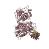
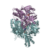
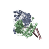
- Assembly
| Deposited unit | 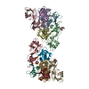
| ||||||||
|---|---|---|---|---|---|---|---|---|---|
| 1 | 
| ||||||||
| 2 | 
| ||||||||
| Unit cell |
| ||||||||
| Atom site foot note | 1: CIS PROLINE - PRO A 196 / 2: CIS PROLINE - PRO D 84 / 3: CIS PROLINE - PRO E 84 / 4: CIS PROLINE - PRO G 196 / 5: CIS PROLINE - PRO J 84 / 6: CIS PROLINE - PRO K 84 | ||||||||
| Noncrystallographic symmetry (NCS) | NCS oper: (Code: given Matrix: (-0.9225, 0.3548, -0.1517), Vector: Details | MTRIX THE TRANSFORMATIONS PRESENTED ON MTRIX RECORDS BELOW DESCRIBE NON-CRYSTALLOGRAPHIC RELATIONSHIPS AMONG THE VARIOUS DOMAINS IN THIS ENTRY. APPLYING THE APPROPRIATE MTRIX TRANSFORMATION TO THE RESIDUES LISTED FIRST WILL YIELD APPROXIMATE COORDINATES FOR THE RESIDUES LISTED SECOND. APPLIED TO TRANSFORMED TO MTRIX RESIDUES RESIDUES RMSD M1 A 2 .. A 235 G 2 .. G 235 0.916 M1 B 4 .. B 199 H 4 .. H 199 0.659 M1 C 4 .. C 199 I 4 .. I 199 0.916 M1 D 1 .. D 110 J 1 .. J 110 0.554 M1 E 1 .. E 110 K 1 .. K 110 1.009 M1 F 2 .. F 99 L 2 .. L 99 0.955 | |
-Components
-PERTUSSIS TOXIN (SUBUNIT ... , 4 types, 9 molecules AGDEJKFLH
| #1: Protein | Mass: 27129.732 Da / Num. of mol.: 2 / Source method: isolated from a natural source / Source: (natural) Bordetella pertussis (bacteria) / Cell line: S2 / References: EMBL: X16347, UniProt: P04977*PLUS#4: Protein | Mass: 12072.426 Da / Num. of mol.: 4 / Source method: isolated from a natural source / Source: (natural) Bordetella pertussis (bacteria) / Cell line: S2 / References: UniProt: P04980, UniProt: P0A3R5*PLUS#5: Protein | Mass: 10894.472 Da / Num. of mol.: 2 / Source method: isolated from a natural source / Source: (natural) Bordetella pertussis (bacteria) / Cell line: S2 / References: UniProt: P04981#6: Protein | | Mass: 21856.625 Da / Num. of mol.: 1 / Source method: isolated from a natural source / Source: (natural) Bordetella pertussis (bacteria) / Cell line: S2 / References: UniProt: P04978 |
|---|
-Protein , 2 types, 3 molecules BCI
| #2: Protein | Mass: 21658.404 Da / Num. of mol.: 1 / Source method: isolated from a natural source Details: SACCHARIDE CONTAINS TERMINAL N-ACETYLNEURAMINIC ACID (ALPHA 2,6) GALACTOSE Source: (natural) Bordetella pertussis (bacteria) / Cell line: S2 / References: UniProt: P04978 |
|---|---|
| #3: Protein | Mass: 21622.543 Da / Num. of mol.: 2 / Source method: isolated from a natural source Details: SACCHARIDE CONTAINS TERMINAL N-ACETYLNEURAMINIC ACID (ALPHA 2,6) GALACTOSE Source: (natural) Bordetella pertussis (bacteria) / Cell line: S2 / References: UniProt: P04979 |
-Sugars , 1 types, 3 molecules
| #7: Polysaccharide | Source method: isolated from a genetically manipulated source |
|---|
-Details
| Has protein modification | Y |
|---|
-Experimental details
-Experiment
| Experiment | Method: X-RAY DIFFRACTION |
|---|
- Sample preparation
| Crystal | Density Matthews: 3.7 Å3/Da / Density % sol: 66.78 % | |||||||||||||||||||||||||||||||||||
|---|---|---|---|---|---|---|---|---|---|---|---|---|---|---|---|---|---|---|---|---|---|---|---|---|---|---|---|---|---|---|---|---|---|---|---|---|
| Crystal grow | *PLUS pH: 8 / Method: vapor diffusion, hanging drop / Details: Stein, P.E., (1994) Structure, 2, 45. | |||||||||||||||||||||||||||||||||||
| Components of the solutions | *PLUS
|
-Data collection
| Radiation | Scattering type: x-ray |
|---|---|
| Radiation wavelength | Relative weight: 1 |
| Reflection | *PLUS Highest resolution: 3.5 Å / Lowest resolution: 10 Å / % possible obs: 87 % / Rmerge(I) obs: 0.097 |
- Processing
| Software |
| ||||||||||||||||||||||||||||||||||||||||||||||||||||||||||||
|---|---|---|---|---|---|---|---|---|---|---|---|---|---|---|---|---|---|---|---|---|---|---|---|---|---|---|---|---|---|---|---|---|---|---|---|---|---|---|---|---|---|---|---|---|---|---|---|---|---|---|---|---|---|---|---|---|---|---|---|---|---|
| Refinement | Resolution: 3.5→10 Å / σ(F): 0 /
| ||||||||||||||||||||||||||||||||||||||||||||||||||||||||||||
| Refinement step | Cycle: LAST / Resolution: 3.5→10 Å
| ||||||||||||||||||||||||||||||||||||||||||||||||||||||||||||
| Refine LS restraints |
| ||||||||||||||||||||||||||||||||||||||||||||||||||||||||||||
| Refinement | *PLUS | ||||||||||||||||||||||||||||||||||||||||||||||||||||||||||||
| Solvent computation | *PLUS | ||||||||||||||||||||||||||||||||||||||||||||||||||||||||||||
| Displacement parameters | *PLUS Biso mean: 20 Å2 |
