 Movie
Movie Controller

[English] 日本語
 Yorodumi
- PDB-6tj2: Extracellular alpha/beta-hydrolase from Paenibacillus species sha... -
+ Open data

- Basic information
| Entry | Database: PDB / ID: 6tj2 | |||||||||
|---|---|---|---|---|---|---|---|---|---|---|
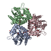
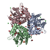
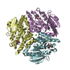
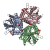
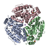
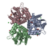
| Title | Extracellular alpha/beta-hydrolase from Paenibacillus species shares structural and functional homology to Tobacco Salicylic Acid Binding Protein 2 | |||||||||
 Components Components | Alpha/beta hydrolase | |||||||||
 Keywords Keywords | HYDROLASE / alpha/beta-hydrolase / methyl salicylate esterase / Paenibacillus / rhizosphere / SABP2 / salicylic acid | |||||||||
| Function / homology | Alpha/beta hydrolase family / Alpha/beta hydrolase fold-1 / Alpha/Beta hydrolase fold / hydrolase activity / Alpha/beta hydrolase Function and homology information Function and homology information | |||||||||
| Biological species |  Paenibacillus sp. VTT E-133280 (bacteria) Paenibacillus sp. VTT E-133280 (bacteria) | |||||||||
| Method |  X-RAY DIFFRACTION / SYNCHROTRON / MOLECULAR REPLACEMENT / Resolution: 1.32 Å X-RAY DIFFRACTION / SYNCHROTRON / MOLECULAR REPLACEMENT / Resolution: 1.32 Å | |||||||||
 Authors Authors | Fulop, V. / Wilkinson, R.C. | |||||||||
| Funding support |  United Kingdom, 2items United Kingdom, 2items
| |||||||||
 Citation Citation | Journal: J.Struct.Biol. / Year: 2020 Title: Extracellular alpha/beta-hydrolase from Paenibacillus species shares structural and functional homology to tobacco salicylic acid binding protein 2. Authors: Wilkinson, R.C. / Rahman Pour, R. / Jamshidi, S. / Fulop, V. / Bugg, T.D.H. | |||||||||
| History |
|
- Structure visualization
| Structure viewer | Molecule: MolmilJmol/JSmol |
|---|
- Downloads & links
-Download
| PDBx/mmCIF format | 6tj2.cif.gz | 167.7 KB | Display | PDBx/mmCIF format |
|---|---|---|---|---|
| PDB format | pdb6tj2.ent.gz | 130.7 KB | Display | PDB format |
| PDBx/mmJSON format | 6tj2.json.gz | Tree view | PDBx/mmJSON format | |
| Others |  Other downloads Other downloads |
-Validation report
| Summary document | 6tj2_validation.pdf.gz | 430.6 KB | Display | wwPDB validaton report |
|---|---|---|---|---|
| Full document | 6tj2_full_validation.pdf.gz | 433.5 KB | Display | |
| Data in XML | 6tj2_validation.xml.gz | 33 KB | Display | |
| Data in CIF | 6tj2_validation.cif.gz | 49.6 KB | Display | |
| Arichive directory | https://data.pdbj.org/pub/pdb/validation_reports/tj/6tj2ftp://data.pdbj.org/pub/pdb/validation_reports/tj/6tj2 | HTTPS FTP |
-Related structure data
| Related structure data | 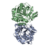 3dqzS S: Starting model for refinement |
|---|---|
| Similar structure data |
-Links
 PDBj
PDBj

- Assembly
| Deposited unit | 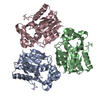
| ||||||||
|---|---|---|---|---|---|---|---|---|---|
| 1 |
| ||||||||
| Unit cell |
|
-Components
| #1: Protein | Mass: 31771.234 Da / Num. of mol.: 3 Source method: isolated from a genetically manipulated source Source: (gene. exp.) Paenibacillus sp. VTT E-133280 (bacteria)Gene: CA600_10415 / Production host: #2: Water | ChemComp-HOH / |  Mass: 18.015 Da / Num. of mol.: 667 / Source method: isolated from a natural source / Formula: H2O Mass: 18.015 Da / Num. of mol.: 667 / Source method: isolated from a natural source / Formula: H2O |
|---|
-Experimental details
-Experiment
| Experiment | Method: X-RAY DIFFRACTION / Number of used crystals: 1 |
|---|
- Sample preparation
| Crystal | Density Matthews: 1.79 Å3/Da / Density % sol: 31.3 % / Description: Rod |
|---|---|
| Crystal grow | Temperature: 295 K / Method: vapor diffusion, sitting drop / Details: 0.2 M sodium fluoride, 20% PEG 3350 |
-Data collection
| Diffraction | Mean temperature: 100 K / Serial crystal experiment: N |
|---|---|
| Diffraction source | Source: SYNCHROTRON / Site: Diamond / Beamline: I03 / Wavelength: 0.91587 Å |
| Detector | Type: DECTRIS PILATUS 6M / Detector: PIXEL / Date: Feb 10, 2017 |
| Radiation | Monochromator: MIRRORS / Protocol: SINGLE WAVELENGTH / Monochromatic (M) / Laue (L): M / Scattering type: x-ray |
| Radiation wavelength | Wavelength: 0.91587 Å / Relative weight: 1 |
| Reflection | Resolution: 1.32→57 Å / Num. obs: 159905 / % possible obs: 99.2 % / Observed criterion σ(I): -3 / Redundancy: 6.5 % / Biso Wilson estimate: 15.2 Å2 / CC1/2: 0.999 / Rmerge(I) obs: 0.059 / Rpim(I) all: 0.025 / Rrim(I) all: 0.064 / Rsym value: 0.059 / Net I/σ(I): 17.7 |
| Reflection shell | Resolution: 1.32→1.39 Å / Redundancy: 6.1 % / Rmerge(I) obs: 0.739 / Mean I/σ(I) obs: 2 / Num. unique obs: 22076 / CC1/2: 0.737 / Rpim(I) all: 0.32 / Rrim(I) all: 0.807 / Rsym value: 0.739 / % possible all: 94.7 |
- Processing
| Software |
| ||||||||||||||||||||||||||||||||||||||||||||||||||||||||||||
|---|---|---|---|---|---|---|---|---|---|---|---|---|---|---|---|---|---|---|---|---|---|---|---|---|---|---|---|---|---|---|---|---|---|---|---|---|---|---|---|---|---|---|---|---|---|---|---|---|---|---|---|---|---|---|---|---|---|---|---|---|---|
| Refinement | Method to determine structure: MOLECULAR REPLACEMENT Starting model: 3DQZ Resolution: 1.32→56.22 Å / Cor.coef. Fo:Fc: 0.973 / Cor.coef. Fo:Fc free: 0.966 / SU B: 0.824 / SU ML: 0.034 / Cross valid method: THROUGHOUT / σ(F): 0 / ESU R: 0.048 / ESU R Free: 0.05 Details: HYDROGENS HAVE BEEN ADDED IN THE RIDING POSITIONS U VALUES : REFINED INDIVIDUALLY
| ||||||||||||||||||||||||||||||||||||||||||||||||||||||||||||
| Solvent computation | Ion probe radii: 0.8 Å / Shrinkage radii: 0.8 Å / VDW probe radii: 1.2 Å | ||||||||||||||||||||||||||||||||||||||||||||||||||||||||||||
| Displacement parameters | Biso max: 79.83 Å2 / Biso mean: 15.128 Å2 / Biso min: 8.63 Å2
| ||||||||||||||||||||||||||||||||||||||||||||||||||||||||||||
| Refinement step | Cycle: final / Resolution: 1.32→56.22 Å
| ||||||||||||||||||||||||||||||||||||||||||||||||||||||||||||
| Refine LS restraints |
| ||||||||||||||||||||||||||||||||||||||||||||||||||||||||||||
| LS refinement shell | Resolution: 1.32→1.354 Å / Rfactor Rfree error: 0 / Total num. of bins used: 20
|
