 Movie
Movie Controller


+ Open data

- Basic information
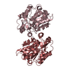
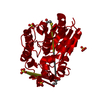
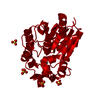
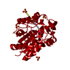
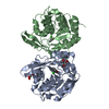
| Entry | Database: PDB / ID: 3dqz | ||||||
|---|---|---|---|---|---|---|---|
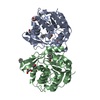
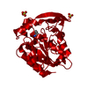
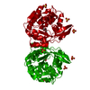
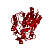
| Title | Structure of the hydroxynitrile lyase from Arabidopsis thaliana | ||||||
 Components Components | Alpha-hydroxynitrile lyase-like protein | ||||||
 Keywords Keywords | LYASE / A/B-Hydrloase fold / Cyanogenesis | ||||||
| Function / homology |  Function and homology information Function and homology information(R)-mandelonitrile lyase / mandelonitrile lyase activity / defense response to insect / methyl salicylate esterase activity / methyl indole-3-acetate esterase activity / methyl jasmonate esterase activity / salicylic acid metabolic process / jasmonic acid metabolic process / glycoside catabolic process / response to wounding ...(R)-mandelonitrile lyase / mandelonitrile lyase activity / defense response to insect / methyl salicylate esterase activity / methyl indole-3-acetate esterase activity / methyl jasmonate esterase activity / salicylic acid metabolic process / jasmonic acid metabolic process / glycoside catabolic process / response to wounding / nucleus / plasma membrane / cytoplasm Similarity search - Function | ||||||
| Biological species |  | ||||||
| Method |  X-RAY DIFFRACTION / SYNCHROTRON / MOLECULAR REPLACEMENT / molecular replacement / Resolution: 2.504 Å X-RAY DIFFRACTION / SYNCHROTRON / MOLECULAR REPLACEMENT / molecular replacement / Resolution: 2.504 Å | ||||||
 Authors Authors | Andexer, J. / Staunig, N. / Gruber, K. | ||||||
 Citation Citation | Journal: Chembiochem / Year: 2012 Title: Hydroxynitrile lyases with alpha / beta-hydrolase fold: two enzymes with almost identical 3D structures but opposite enantioselectivities and different reaction mechanisms Authors: Andexer, J.N. / Staunig, N. / Eggert, T. / Kratky, C. / Pohl, M. / Gruber, K. | ||||||
| History |
|
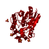
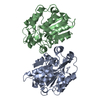
- Structure visualization
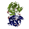
| Structure viewer | Molecule: MolmilJmol/JSmol |
|---|
- Downloads & links
-Download
| PDBx/mmCIF format | 3dqz.cif.gz | 205.2 KB | Display | PDBx/mmCIF format |
|---|---|---|---|---|
| PDB format | pdb3dqz.ent.gz | 165.6 KB | Display | PDB format |
| PDBx/mmJSON format | 3dqz.json.gz | Tree view | PDBx/mmJSON format | |
| Others |  Other downloads Other downloads |
-Validation report
| Arichive directory | https://data.pdbj.org/pub/pdb/validation_reports/dq/3dqzftp://data.pdbj.org/pub/pdb/validation_reports/dq/3dqz | HTTPS FTP |
|---|
-Related structure data


-Links
 PDBj
PDBj


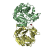
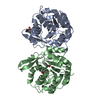
- Assembly
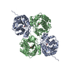
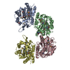
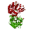
| Deposited unit | 
| ||||||||||||||||
|---|---|---|---|---|---|---|---|---|---|---|---|---|---|---|---|---|---|
| 1 | 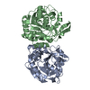
| ||||||||||||||||
| 2 | 
| ||||||||||||||||
| Unit cell |
| ||||||||||||||||
| Noncrystallographic symmetry (NCS) | NCS oper:
|
-Components
| #1: Protein | Mass: 29253.684 Da / Num. of mol.: 4 Source method: isolated from a genetically manipulated source Source: (gene. exp.)  #2: Chemical | ChemComp-CL / |   Mass: 35.453 Da / Num. of mol.: 1 / Source method: obtained synthetically / Formula: Cl Mass: 35.453 Da / Num. of mol.: 1 / Source method: obtained synthetically / Formula: Cl#3: Water | ChemComp-HOH / |  Mass: 18.015 Da / Num. of mol.: 69 / Source method: isolated from a natural source / Formula: H2O Mass: 18.015 Da / Num. of mol.: 69 / Source method: isolated from a natural source / Formula: H2O |
|---|
-Experimental details
-Experiment
| Experiment | Method: X-RAY DIFFRACTION / Number of used crystals: 1 |
|---|
- Sample preparation
| Crystal | Density Matthews: 2.36 Å3/Da / Density % sol: 47.86 % / Mosaicity: 0.834 ° |
|---|---|
| Crystal grow | Temperature: 293 K / Method: vapor diffusion, sitting drop / pH: 6 Details: 10-18% PEG3350, 100mM BisTris, pH6.0, VAPOR DIFFUSION, SITTING DROP, temperature 293K |
-Data collection
| Diffraction | Mean temperature: 100 K |
|---|---|
| Diffraction source | Source: SYNCHROTRON / Site: EMBL/DESY, HAMBURG  / Beamline: X13 / Wavelength: 0.8081 Å / Beamline: X13 / Wavelength: 0.8081 Å |
| Detector | Type: MAR CCD 165 mm / Detector: CCD / Date: Jun 15, 2007 |
| Radiation | Protocol: SINGLE WAVELENGTH / Monochromatic (M) / Laue (L): M / Scattering type: x-ray |
| Radiation wavelength | Wavelength: 0.8081 Å / Relative weight: 1 |
| Reflection twin | Operator: l,-k,h / Fraction: 0.481 |
| Reflection | Resolution: 2.5→30 Å / Num. all: 33151 / Num. obs: 33151 / % possible obs: 89.6 % / Redundancy: 3.4 % / Biso Wilson estimate: 28.3 Å2 / Rsym value: 0.071 / Χ2: 1.076 / Net I/σ(I): 18.9 |
| Reflection shell | Resolution: 2.5→2.56 Å / Redundancy: 2.9 % / Mean I/σ(I) obs: 4.8 / Num. unique all: 2107 / Rsym value: 0.21 / Χ2: 0.939 / % possible all: 87.5 |
-Phasing
| Phasing | Method: molecular replacement |
|---|
- Processing
| Software |
| ||||||||||||||||||||||||||||
|---|---|---|---|---|---|---|---|---|---|---|---|---|---|---|---|---|---|---|---|---|---|---|---|---|---|---|---|---|---|
| Refinement | Method to determine structure: MOLECULAR REPLACEMENT Starting model: PDB ENTRY 1QJ4, 1DWP, 1XKL Resolution: 2.504→24.171 Å / Occupancy max: 1 / Occupancy min: 1 / FOM work R set: 0.862 / Isotropic thermal model: isotropic / Cross valid method: THROUGHOUT / Stereochemistry target values: Engh & Huber Details: This is a twinned structure, the detwin fraction is 0.481 and operator is 'l,-k,h'.
| ||||||||||||||||||||||||||||
| Solvent computation | Shrinkage radii: 0.9 Å / VDW probe radii: 1.11 Å / Solvent model: FLAT BULK SOLVENT MODEL / Bsol: 24.792 Å2 / ksol: 0.352 e/Å3 | ||||||||||||||||||||||||||||
| Displacement parameters | Biso max: 86.9 Å2 / Biso mean: 27.936 Å2 / Biso min: 2.78 Å2
| ||||||||||||||||||||||||||||
| Refinement step | Cycle: LAST / Resolution: 2.504→24.171 Å
| ||||||||||||||||||||||||||||
| Refine LS restraints |
|
