 Movie
Movie Controller


+ Open data

- Basic information
| Entry | Database: PDB / ID: 3c6z | ||||||
|---|---|---|---|---|---|---|---|
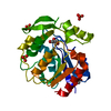
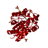
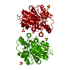
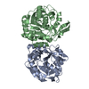
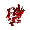
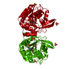
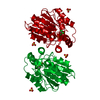
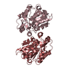
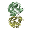
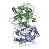
| Title | HNL from Hevea brasiliensis to atomic resolution | ||||||
 Components Components | Hydroxynitrilase | ||||||
 Keywords Keywords | LYASE / atomic resolution / hydroxynitril lyase / catalysis / protonation state / ab initio calculations / substrate binding | ||||||
| Function / homology |  Function and homology information Function and homology informationaliphatic (S)-hydroxynitrile lyase activity / aromatic (S)-hydroxynitrile lyase activity / (S)-hydroxynitrile lyase / methyl salicylate esterase activity / methyl indole-3-acetate esterase activity / methyl jasmonate esterase activity / salicylic acid metabolic process / jasmonic acid metabolic process Similarity search - Function | ||||||
| Biological species |   Hevea brasiliensis (rubber tree) Hevea brasiliensis (rubber tree) | ||||||
| Method |  X-RAY DIFFRACTION / SYNCHROTRON / molecular replacement/rigid body placement / Resolution: 1.05 Å X-RAY DIFFRACTION / SYNCHROTRON / molecular replacement/rigid body placement / Resolution: 1.05 Å | ||||||
 Authors Authors | Schmidt, A. | ||||||
 Citation Citation | Journal: J.Biol.Chem. / Year: 2008 Title: Atomic resolution crystal structures and quantum chemistry meet to reveal subtleties of hydroxynitrile lyase catalysis Authors: Schmidt, A. / Gruber, K. / Kratky, C. / Lamzin, V.S. #1: Journal: Protein Sci. / Year: 1999Title: Three-dimensional structures of enzyme-substrate complexes of the hydroxynitrile lyase from Hevea brasiliensis Authors: Zuegg, J. / Gruber, K. / Gugganig, M. / Wagner, U.G. / Kratky, C. #2: Journal: Biol.Chem. / Year: 1999Title: Atomic resolution crystal structure of hydroxynitrile lyase from Hevea brasiliensis Authors: Gruber, K. / Gugganig, M. / Wagner, U.G. / Kratky, C. | ||||||
| History |
|
- Structure visualization
| Structure viewer | Molecule: MolmilJmol/JSmol |
|---|
- Downloads & links
-Download
| PDBx/mmCIF format | 3c6z.cif.gz | 196.8 KB | Display | PDBx/mmCIF format |
|---|---|---|---|---|
| PDB format | pdb3c6z.ent.gz | 159.7 KB | Display | PDB format |
| PDBx/mmJSON format | 3c6z.json.gz | Tree view | PDBx/mmJSON format | |
| Others |  Other downloads Other downloads |
-Validation report
| Arichive directory | https://data.pdbj.org/pub/pdb/validation_reports/c6/3c6zftp://data.pdbj.org/pub/pdb/validation_reports/c6/3c6z | HTTPS FTP |
|---|
-Related structure data
| Related structure data |  3c6xC  3c6yC  3c70C 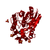 1qj4S S: Starting model for refinement C: citing same article ( |
|---|---|
| Similar structure data |

-Links
 PDBj
PDBj


- Assembly
| Deposited unit | 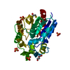
| |||||||||||||||||||||
|---|---|---|---|---|---|---|---|---|---|---|---|---|---|---|---|---|---|---|---|---|---|---|
| 1 | 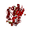
| |||||||||||||||||||||
| Unit cell |
| |||||||||||||||||||||
| Components on special symmetry positions |
|
-Components
-Protein , 1 types, 1 molecules A
| #1: Protein | Mass: 29262.598 Da / Num. of mol.: 1 Source method: isolated from a genetically manipulated source Source: (gene. exp.) Hevea brasiliensis (rubber tree) / Gene: HNL / Plasmid: BHIL-D2 / Production host:  Pichia pastoris (fungus) / References: UniProt: P52704, EC: 4.1.2.37 Pichia pastoris (fungus) / References: UniProt: P52704, EC: 4.1.2.37 |
|---|
-Non-polymers , 5 types, 615 molecules 


| #2: Chemical | ChemComp-SO4 /  Mass: 96.063 Da / Num. of mol.: 4 / Source method: obtained synthetically / Formula: SO4 Mass: 96.063 Da / Num. of mol.: 4 / Source method: obtained synthetically / Formula: SO4#3: Chemical | ChemComp-BME / |  Mass: 78.133 Da / Num. of mol.: 1 / Source method: obtained synthetically / Formula: C2H6OS Mass: 78.133 Da / Num. of mol.: 1 / Source method: obtained synthetically / Formula: C2H6OS#4: Chemical | ChemComp-IPA / |  Mass: 60.095 Da / Num. of mol.: 1 / Source method: obtained synthetically / Formula: C3H8O / Comment: alkaloid*YM Mass: 60.095 Da / Num. of mol.: 1 / Source method: obtained synthetically / Formula: C3H8O / Comment: alkaloid*YM#5: Chemical | ChemComp-PEG / |  Mass: 106.120 Da / Num. of mol.: 1 / Source method: obtained synthetically / Formula: C4H10O3 Mass: 106.120 Da / Num. of mol.: 1 / Source method: obtained synthetically / Formula: C4H10O3#6: Water | ChemComp-HOH / | Mass: 18.015 Da / Num. of mol.: 608 / Source method: isolated from a natural source / Formula: H2O |
|---|
-Experimental details
-Experiment
| Experiment | Method: X-RAY DIFFRACTION / Number of used crystals: 1 |
|---|
- Sample preparation
| Crystal | Density Matthews: 2.75 Å3/Da / Density % sol: 55.34 % |
|---|---|
| Crystal grow | Temperature: 293 K / Method: vapor diffusion, hanging drop / pH: 7.5 Details: 2% PEG400, 2M ammonium sulphate, 0.1M Hepes-Na, pH7.5, VAPOR DIFFUSION, HANGING DROP, temperature 293K |
-Data collection
| Diffraction | Mean temperature: 100 K |
|---|---|
| Diffraction source | Source: SYNCHROTRON / Site: EMBL/DESY, HAMBURG  / Beamline: BW7B / Wavelength: 0.843 Å / Beamline: BW7B / Wavelength: 0.843 Å |
| Detector | Type: MAR scanner 345 mm plate / Detector: IMAGE PLATE / Date: Mar 6, 2005 / Details: mirror |
| Radiation | Monochromator: Si / Protocol: SINGLE WAVELENGTH / Monochromatic (M) / Laue (L): M / Scattering type: x-ray |
| Radiation wavelength | Wavelength: 0.843 Å / Relative weight: 1 |
| Reflection | Resolution: 1.05→30 Å / Num. all: 140831 / Num. obs: 140831 / % possible obs: 100 % / Observed criterion σ(F): -3 / Observed criterion σ(I): -3 / Redundancy: 3.9 % / Biso Wilson estimate: 8.2 Å2 / Rmerge(I) obs: 0.046 / Net I/σ(I): 26 |
| Reflection shell | Resolution: 1.05→1.06 Å / Redundancy: 2 % / Rmerge(I) obs: 0.29 / Mean I/σ(I) obs: 3.1 / Num. unique all: 1721 / % possible all: 50 |
- Processing
| Software |
| |||||||||||||||||||||||||||||||||
|---|---|---|---|---|---|---|---|---|---|---|---|---|---|---|---|---|---|---|---|---|---|---|---|---|---|---|---|---|---|---|---|---|---|---|
| Refinement | Method to determine structure: molecular replacement/rigid body placement Starting model: PDB ENTRY 1QJ4 Resolution: 1.05→30 Å / Num. parameters: 25764 / Num. restraintsaints: 31662 Cross valid method: FREE R initially, final refinement against all data σ(F): 0 / σ(I): 0 / Stereochemistry target values: ENGH AND HUBER / Details: full anisotropic refinement applied
| |||||||||||||||||||||||||||||||||
| Displacement parameters | Biso mean: 11 Å2 | |||||||||||||||||||||||||||||||||
| Refine analyze | Num. disordered residues: 38 / Occupancy sum hydrogen: 2040.7 / Occupancy sum non hydrogen: 2654.25 | |||||||||||||||||||||||||||||||||
| Refinement step | Cycle: LAST / Resolution: 1.05→30 Å
| |||||||||||||||||||||||||||||||||
| Refine LS restraints |
|
