 Movie
Movie Controller

[English] 日本語
 Yorodumi
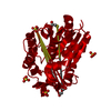
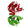
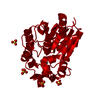
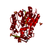
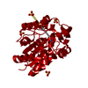
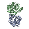
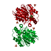
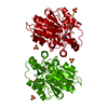
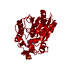
- PDB-1qj4: HYDROXYNITRILE-LYASE FROM HEVEA BRASILIENSIS AT ATOMIC RESOLUTION -
+ Open data

- Basic information
| Entry | Database: PDB / ID: 1qj4 | ||||||
|---|---|---|---|---|---|---|---|
| Title | HYDROXYNITRILE-LYASE FROM HEVEA BRASILIENSIS AT ATOMIC RESOLUTION | ||||||
 Components Components | HYDROXYNITRILE LYASE | ||||||
 Keywords Keywords | LYASE / OXYNITRILASE / CYANOGENESIS / CYANHYDRIN FORMATION | ||||||
| Function / homology |  Function and homology information Function and homology informationaliphatic (S)-hydroxynitrile lyase activity / aromatic (S)-hydroxynitrile lyase activity / (S)-hydroxynitrile lyase / methyl salicylate esterase activity / methyl indole-3-acetate esterase activity / methyl jasmonate esterase activity / salicylic acid metabolic process / jasmonic acid metabolic process Similarity search - Function | ||||||
| Biological species |   HEVEA BRASILIENSIS (rubber tree) HEVEA BRASILIENSIS (rubber tree) | ||||||
| Method |  X-RAY DIFFRACTION / SYNCHROTRON / MOLECULAR REPLACEMENT / Resolution: 1.1 Å X-RAY DIFFRACTION / SYNCHROTRON / MOLECULAR REPLACEMENT / Resolution: 1.1 Å | ||||||
 Authors Authors | Gugganig, M. / Gruber, K. / Kratky, C. | ||||||
 Citation Citation | Journal: Biol.Chem. / Year: 1999 Title: Atomic Resolution Crystal Structure of Hydroxynitrile Lyase from Hevea Brasiliensis Authors: Gruber, K. / Gugganig, M. / Wagner, U.G. / Kratky, C. #1: Journal: Protein Sci. / Year: 2000Title: Three-Dimensional Structures of Enzyme-Substrate Complexes of the Hydroxynitrile Lyase from Hevea Brasiliensis Authors: Zuegg, J. / Gruber, K. / Gugganig, M. / Wagner, U.G. / Kratky, C. #2: Journal: Structure / Year: 1996Title: Mechanism of Cyanogenesis: The Crystal Structure of Hydroxynitrile Lyase from Hevea Brasiliensis Authors: Wagner, U.G. / Hasslacher, M. / Griengl, H. / Schwab, H. / Kratky, C. #3: Journal: Acta Crystallogr.,Sect.D / Year: 1996 Title: Crystallization of a Hydroxynitrile Lyase Authors: Wagner, U.G. / Schall, M. / Hayn, M. / Hasslacher, M. / Schwab, H. / Griengl, H.S. / Kratky, C. #4: Journal: J.Biol.Chem. / Year: 1996 Title: Molecular Cloning of the Full-Length Cdna of (S)-Hydroxynitrile Lyase from Hevea Brasiliensis. Functional Expression in Escherichia Coli and Saccharomyces Cerevisiae and Identification of an Active Site Residue Authors: Hasslacher, M. / Schall, M. / Hayn, M. / Griengl, H. / Kohlwein, S.D. / Schwab, H. | ||||||
| History |
| ||||||
| Remark 650 | HELIX DETERMINATION METHOD: DSSP | ||||||
| Remark 700 | SHEET DETERMINATION METHOD: DSSP |
- Structure visualization
| Structure viewer | Molecule: MolmilJmol/JSmol |
|---|
- Downloads & links
-Download
| PDBx/mmCIF format | 1qj4.cif.gz | 144.3 KB | Display | PDBx/mmCIF format |
|---|---|---|---|---|
| PDB format | pdb1qj4.ent.gz | 114 KB | Display | PDB format |
| PDBx/mmJSON format | 1qj4.json.gz | Tree view | PDBx/mmJSON format | |
| Others |  Other downloads Other downloads |
-Validation report
| Arichive directory | https://data.pdbj.org/pub/pdb/validation_reports/qj/1qj4ftp://data.pdbj.org/pub/pdb/validation_reports/qj/1qj4 | HTTPS FTP |
|---|
-Related structure data
| Related structure data | 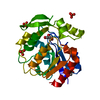 7yasS S: Starting model for refinement |
|---|---|
| Similar structure data |

-Links
 PDBj
PDBj


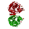
- Assembly
| Deposited unit | 
| |||||||||||||||||||||
|---|---|---|---|---|---|---|---|---|---|---|---|---|---|---|---|---|---|---|---|---|---|---|
| 1 | 
| |||||||||||||||||||||
| Unit cell |
| |||||||||||||||||||||
| Components on special symmetry positions |
|
-Components
| #1: Protein | Mass: 29262.598 Da / Num. of mol.: 1 Source method: isolated from a genetically manipulated source Source: (gene. exp.) HEVEA BRASILIENSIS (rubber tree) / Gene: HNL / Organ: LEAF / Plasmid: BHIL-D2 / Gene (production host): HNL / Production host:  PICHIA PASTORIS (fungus) / References: UniProt: P52704, EC: 4.1.2.39 PICHIA PASTORIS (fungus) / References: UniProt: P52704, EC: 4.1.2.39 | ||
|---|---|---|---|
| #2: Chemical | ChemComp-GOL /   Mass: 92.094 Da / Num. of mol.: 1 / Source method: obtained synthetically / Formula: C3H8O3 Mass: 92.094 Da / Num. of mol.: 1 / Source method: obtained synthetically / Formula: C3H8O3 | ||
| #3: Chemical | ChemComp-SO4 /   Mass: 96.063 Da / Num. of mol.: 4 / Source method: obtained synthetically / Formula: SO4 Mass: 96.063 Da / Num. of mol.: 4 / Source method: obtained synthetically / Formula: SO4#4: Water | ChemComp-HOH / |  Mass: 18.015 Da / Num. of mol.: 550 / Source method: isolated from a natural source / Formula: H2O Mass: 18.015 Da / Num. of mol.: 550 / Source method: isolated from a natural source / Formula: H2O |
-Experimental details
-Experiment
| Experiment | Method: X-RAY DIFFRACTION / Number of used crystals: 1 |
|---|
- Sample preparation
| Crystal | Density Matthews: 2.8 Å3/Da / Density % sol: 56 % |
|---|---|
| Crystal grow | pH: 7.4 Details: 0.1 M NATRIUM HEPES BUFFER PH=7.4 2 % PEG 400, 2.0 M AMMONIUM SULFATE, ROOM-TEMP., pH 7.40 |
-Data collection
| Diffraction | Mean temperature: 120 K |
|---|---|
| Diffraction source | Source: SYNCHROTRON / Site: EMBL/DESY, HAMBURG  / Beamline: BW7B / Wavelength: 0.8345 / Beamline: BW7B / Wavelength: 0.8345 |
| Detector | Type: MARRESEARCH / Detector: IMAGE PLATE / Date: Aug 15, 1998 |
| Radiation | Protocol: SINGLE WAVELENGTH / Monochromatic (M) / Laue (L): M / Scattering type: x-ray |
| Radiation wavelength | Wavelength: 0.8345 Å / Relative weight: 1 |
| Reflection | Resolution: 1.1→50 Å / Num. obs: 123339 / % possible obs: 94.1 % / Observed criterion σ(I): 0 / Redundancy: 4.1 % / Rsym value: 0.065 / Net I/σ(I): 20.4 |
| Reflection shell | Resolution: 1.1→1.11 Å / Redundancy: 3.8 % / Mean I/σ(I) obs: 3 / Rsym value: 0.364 / % possible all: 92.3 |
- Processing
| Software |
| |||||||||||||||||||||||||||||||||
|---|---|---|---|---|---|---|---|---|---|---|---|---|---|---|---|---|---|---|---|---|---|---|---|---|---|---|---|---|---|---|---|---|---|---|
| Refinement | Method to determine structure: MOLECULAR REPLACEMENT Starting model: 7YAS Resolution: 1.1→50 Å / Num. parameters: 24572 / Num. restraintsaints: 43482 / Cross valid method: FREE R-VALUE / σ(F): 0 / Stereochemistry target values: ENGH AND HUBER Details: ANISOTROPIC REFINEMENT REDUCED FREE R (NO CUTOFF) BY 0.043. RESIDUE SER-A80 IS OBSERVED IN A RAMACHANDRAN 'FORBIDDEN' REGION. THIS CONFORMATION IS FOUND IN ALL PREVIOUS HNL-STRUCTURES. ...Details: ANISOTROPIC REFINEMENT REDUCED FREE R (NO CUTOFF) BY 0.043. RESIDUE SER-A80 IS OBSERVED IN A RAMACHANDRAN 'FORBIDDEN' REGION. THIS CONFORMATION IS FOUND IN ALL PREVIOUS HNL-STRUCTURES. STRUCTURES OF OTHER MEMBERS OF THE ALPHA/BETA HYDROLASES FAMILY ALSO CONSISTENTLY SHOW THE EQUIVALENT RESIDUE (NUCLEOPHILE) IN SUCH A CONFORMATION.
| |||||||||||||||||||||||||||||||||
| Solvent computation | Solvent model: MOEWS & KRETSINGER, J.MOL.BIOL.91(1973)201-2 | |||||||||||||||||||||||||||||||||
| Refine analyze | Num. disordered residues: 25 / Occupancy sum hydrogen: 2053 / Occupancy sum non hydrogen: 2629.75 | |||||||||||||||||||||||||||||||||
| Refinement step | Cycle: LAST / Resolution: 1.1→50 Å
| |||||||||||||||||||||||||||||||||
| Refine LS restraints |
|
