 Movie
Movie Controller

[English] 日本語
 Yorodumi
- PDB-3fdu: Crystal structure of a putative enoyl-CoA hydratase/isomerase fro... -
+ Open data

- Basic information
| Entry | Database: PDB / ID: 3fdu | ||||||
|---|---|---|---|---|---|---|---|
| Title | Crystal structure of a putative enoyl-CoA hydratase/isomerase from Acinetobacter baumannii | ||||||
 Components Components | Putative enoyl-CoA hydratase/isomerase | ||||||
 Keywords Keywords | ISOMERASE / STRUCTURAL GENOMICS / PSI-2 / Protein Structure Initiative / New York SGX Research Center for Structural Genomics / NYSGXRC | ||||||
| Function / homology | Lyase 2-enoyl-coa Hydratase, Chain A, domain 2 / Lyase 2-enoyl-coa Hydratase; Chain A, domain 2 / 2-enoyl-CoA Hydratase; Chain A, domain 1 / 2-enoyl-CoA Hydratase; Chain A, domain 1 / Alpha-Beta Complex / Orthogonal Bundle / Mainly Alpha / Alpha Beta / :  Function and homology information Function and homology information | ||||||
| Biological species |  Acinetobacter baumannii (bacteria) Acinetobacter baumannii (bacteria) | ||||||
| Method |  X-RAY DIFFRACTION / SYNCHROTRON / SAD / Resolution: 2 Å X-RAY DIFFRACTION / SYNCHROTRON / SAD / Resolution: 2 Å | ||||||
 Authors Authors | Bonanno, J.B. / Dickey, M. / Bain, K.T. / Tang, B.K. / Romero, R. / Wasserman, S. / Sauder, J.M. / Burley, S.K. / Almo, S.C. / New York SGX Research Center for Structural Genomics (NYSGXRC) | ||||||
 Citation Citation | Journal: To be Published Title: Crystal structure of a putative enoyl-CoA hydratase/isomerase from Acinetobacter baumannii Authors: Bonanno, J.B. / Dickey, M. / Bain, K.T. / Tang, B.K. / Romero, R. / Wasserman, S. / Sauder, J.M. / Burley, S.K. / Almo, S.C. | ||||||
| History |
|
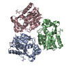
- Structure visualization
| Structure viewer | Molecule: MolmilJmol/JSmol |
|---|
- Downloads & links
-Download
| PDBx/mmCIF format | 3fdu.cif.gz | 280.2 KB | Display | PDBx/mmCIF format |
|---|---|---|---|---|
| PDB format | pdb3fdu.ent.gz | 225.6 KB | Display | PDB format |
| PDBx/mmJSON format | 3fdu.json.gz | Tree view | PDBx/mmJSON format | |
| Others |  Other downloads Other downloads |
-Validation report
| Arichive directory | https://data.pdbj.org/pub/pdb/validation_reports/fd/3fduftp://data.pdbj.org/pub/pdb/validation_reports/fd/3fdu | HTTPS FTP |
|---|
-Related structure data
| Similar structure data | |
|---|---|
| Other databases |


-Links
 PDBj
PDBj- Assembly
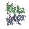
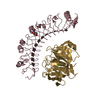
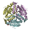
| Deposited unit | 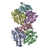
| ||||||||
|---|---|---|---|---|---|---|---|---|---|
| 1 | 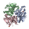
| ||||||||
| 2 | 
| ||||||||
| Unit cell |
| ||||||||
| Details | trimer |
-Components
| #1: Protein | Mass: 28994.230 Da / Num. of mol.: 6 / Mutation: Q11P, Q14N, I59V, K259Q Source method: isolated from a genetically manipulated source Source: (gene. exp.) Acinetobacter baumannii (bacteria) / Strain: ATCC 17978 / Gene: A1S_2548 / Plasmid: modified pET26 / Production host: #2: Chemical | ChemComp-SO4 /   Mass: 96.063 Da / Num. of mol.: 4 / Source method: obtained synthetically / Formula: SO4 Mass: 96.063 Da / Num. of mol.: 4 / Source method: obtained synthetically / Formula: SO4#3: Chemical | ChemComp-GOL / |   Mass: 92.094 Da / Num. of mol.: 1 / Source method: obtained synthetically / Formula: C3H8O3 Mass: 92.094 Da / Num. of mol.: 1 / Source method: obtained synthetically / Formula: C3H8O3#4: Water | ChemComp-HOH / |  Mass: 18.015 Da / Num. of mol.: 683 / Source method: isolated from a natural source / Formula: H2O Mass: 18.015 Da / Num. of mol.: 683 / Source method: isolated from a natural source / Formula: H2O |
|---|
-Experimental details
-Experiment
| Experiment | Method: X-RAY DIFFRACTION / Number of used crystals: 1 |
|---|
- Sample preparation
| Crystal | Density Matthews: 2.32 Å3/Da / Density % sol: 46.91 % |
|---|---|
| Crystal grow | Temperature: 294 K / Method: vapor diffusion / pH: 5 Details: 100mM Bis-Tris pH 5.0, 23% PEG 3350, 200mM ammonium sulfate, vapor diffusion, temperature 294K |
-Data collection
| Diffraction | Mean temperature: 100 K |
|---|---|
| Diffraction source | Source: SYNCHROTRON / Site: APS  / Beamline: 31-ID / Wavelength: 0.97958 Å / Beamline: 31-ID / Wavelength: 0.97958 Å |
| Detector | Type: MAR CCD 165 mm / Detector: CCD / Date: Nov 19, 2008 |
| Radiation | Monochromator: diamond / Protocol: SINGLE WAVELENGTH / Monochromatic (M) / Laue (L): M / Scattering type: x-ray |
| Radiation wavelength | Wavelength: 0.97958 Å / Relative weight: 1 |
| Reflection | Resolution: 2→42.563 Å / Num. all: 107766 / Num. obs: 106796 / % possible obs: 99.1 % / Observed criterion σ(F): 0 / Observed criterion σ(I): 0 / Redundancy: 5.5 % / Biso Wilson estimate: 26.4 Å2 / Rmerge(I) obs: 0.077 / Rsym value: 0.077 / Net I/σ(I): 12.8 |
| Reflection shell | Resolution: 2→2.11 Å / Redundancy: 5.4 % / Rmerge(I) obs: 0.302 / Mean I/σ(I) obs: 4.5 / Num. measured all: 83491 / Num. unique all: 15347 / Rsym value: 0.302 / % possible all: 97.8 |
- Processing
| Software |
| |||||||||||||||||||||||||||||||||||||||||||||||||||||||||||||||||
|---|---|---|---|---|---|---|---|---|---|---|---|---|---|---|---|---|---|---|---|---|---|---|---|---|---|---|---|---|---|---|---|---|---|---|---|---|---|---|---|---|---|---|---|---|---|---|---|---|---|---|---|---|---|---|---|---|---|---|---|---|---|---|---|---|---|---|
| Refinement | Method to determine structure: SAD / Resolution: 2→20 Å / Cor.coef. Fo:Fc: 0.934 / Cor.coef. Fo:Fc free: 0.902 / WRfactor Rfree: 1.083 / WRfactor Rwork: 0.983 / Occupancy max: 1 / Occupancy min: 0.5 / FOM work R set: 0.768 / SU B: 4.581 / SU ML: 0.13 / SU R Cruickshank DPI: 0.204 / SU Rfree: 0.191 / Cross valid method: THROUGHOUT / σ(F): 0 / σ(I): 0 / ESU R: 0.204 / ESU R Free: 0.191 / Stereochemistry target values: MAXIMUM LIKELIHOOD
| |||||||||||||||||||||||||||||||||||||||||||||||||||||||||||||||||
| Solvent computation | Ion probe radii: 0.8 Å / Shrinkage radii: 0.8 Å / VDW probe radii: 1.2 Å / Solvent model: BABINET MODEL WITH MASK | |||||||||||||||||||||||||||||||||||||||||||||||||||||||||||||||||
| Displacement parameters | Biso max: 76.48 Å2 / Biso mean: 33.189 Å2 / Biso min: 11.72 Å2
| |||||||||||||||||||||||||||||||||||||||||||||||||||||||||||||||||
| Refinement step | Cycle: LAST / Resolution: 2→20 Å
| |||||||||||||||||||||||||||||||||||||||||||||||||||||||||||||||||
| Refine LS restraints |
| |||||||||||||||||||||||||||||||||||||||||||||||||||||||||||||||||
| LS refinement shell | Resolution: 2→2.051 Å / Total num. of bins used: 20
|
