 Movie
Movie Controller

[English] 日本語
 Yorodumi
- PDB-6t21: N-terminal domain of EcoKMcrA restriction endonuclease (NEco) in ... -
+ Open data

- Basic information
| Entry | Database: PDB / ID: 6t21 | ||||||
|---|---|---|---|---|---|---|---|
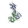
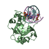
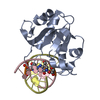
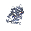
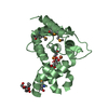
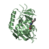
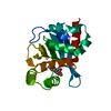
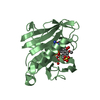
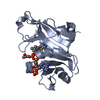
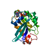
| Title | N-terminal domain of EcoKMcrA restriction endonuclease (NEco) in complex with T5mCGA target sequence | ||||||
 Components Components |
| ||||||
 Keywords Keywords | HYDROLASE / EcoKMcrA / NEco / N-TERMINAL DOMAIN / MODIFICATION DEPENDENT RESTRICTION / 5-METHYLCYTOSINE / 5MC / 5-HYDROXYMETHYLCYTOSINE / 5HMC / HNH ENDONUCLEASE / BBA-ME NUCLEASE | ||||||
| Function / homology |  Function and homology information Function and homology informationHydrolases; Acting on ester bonds; Endodeoxyribonucleases producing 5'-phosphomonoesters / DNA restriction-modification system / methyl-CpG binding / endonuclease activity / hydrolase activity / zinc ion binding Similarity search - Function | ||||||
| Biological species |  synthetic construct (others) | ||||||
| Method |  X-RAY DIFFRACTION / SYNCHROTRON / MOLECULAR REPLACEMENT / Resolution: 2.07 Å X-RAY DIFFRACTION / SYNCHROTRON / MOLECULAR REPLACEMENT / Resolution: 2.07 Å | ||||||
 Authors Authors | Slyvka, A. / Zagorskaite, E. / Czapinska, H. / Sasnauskas, G. / Bochtler, M. | ||||||
 Citation Citation | Journal: Nucleic Acids Res. / Year: 2019 Title: Crystal structure of the EcoKMcrA N-terminal domain (NEco): recognition of modified cytosine bases without flipping. Authors: Slyvka, A. / Zagorskaite, E. / Czapinska, H. / Sasnauskas, G. / Bochtler, M. #1: Journal: Nucleic Acids Res. / Year: 2018Title: Activity and structure of EcoKMcrA. Authors: Czapinska, H. / Kowalska, M. / Zagorskaite, E. / Manakova, E. / Slyvka, A. / Xu, S.Y. / Siksnys, V. / Sasnauskas, G. / Bochtler, M. | ||||||
| History |
|
- Structure visualization
| Structure viewer | Molecule: MolmilJmol/JSmol |
|---|
- Downloads & links
-Download
| PDBx/mmCIF format | 6t21.cif.gz | 191.2 KB | Display | PDBx/mmCIF format |
|---|---|---|---|---|
| PDB format | pdb6t21.ent.gz | 149 KB | Display | PDB format |
| PDBx/mmJSON format | 6t21.json.gz | Tree view | PDBx/mmJSON format | |
| Others |  Other downloads Other downloads |
-Validation report
| Arichive directory | https://data.pdbj.org/pub/pdb/validation_reports/t2/6t21ftp://data.pdbj.org/pub/pdb/validation_reports/t2/6t21 | HTTPS FTP |
|---|
-Related structure data
| Related structure data | 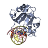 6r64SC  6t22C S: Starting model for refinement C: citing same article ( |
|---|---|
| Similar structure data |

-Links
 PDBj
PDBj
- Assembly
| Deposited unit | 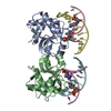
| ||||||||
|---|---|---|---|---|---|---|---|---|---|
| 1 | 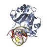
| ||||||||
| 2 | 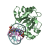
| ||||||||
| Unit cell |
| ||||||||
| Components on special symmetry positions |
|
-Components
| #1: Protein | Mass: 17368.604 Da / Num. of mol.: 2 Source method: isolated from a genetically manipulated source Source: (gene. exp.) Strain: K12 / Gene: mcrA, rglA, b1159, JW1145 / Plasmid: PET15BM / Production host: References: UniProt: P24200, Hydrolases; Acting on ester bonds; Endodeoxyribonucleases producing 5'-phosphomonoesters #2: DNA chain | Mass: 3009.006 Da / Num. of mol.: 2 Source method: isolated from a genetically manipulated source Source: (gene. exp.) synthetic construct (others) / Production host: synthetic construct (others) #3: DNA chain | Mass: 3107.081 Da / Num. of mol.: 2 Source method: isolated from a genetically manipulated source Source: (gene. exp.) synthetic construct (others) / Production host: synthetic construct (others) #4: Water | ChemComp-HOH / |  Mass: 18.015 Da / Num. of mol.: 431 / Source method: isolated from a natural source / Formula: H2O Mass: 18.015 Da / Num. of mol.: 431 / Source method: isolated from a natural source / Formula: H2OHas ligand of interest | Y | |
|---|
-Experimental details
-Experiment
| Experiment | Method: X-RAY DIFFRACTION / Number of used crystals: 1 |
|---|
- Sample preparation
| Crystal | Density Matthews: 3.05 Å3/Da / Density % sol: 59.66 % |
|---|---|
| Crystal grow | Temperature: 291 K / Method: vapor diffusion, hanging drop / pH: 6.5 Details: 1.8 ul of protein-DNA solution (436:523 uM) was mixed with 2.2 ul of the F1 condition of the PACT premier crystal screen (MDL) (0.2 M Sodium fluoride, 0.1 M Bis-Tris propane, pH 6.5, 20% PEG ...Details: 1.8 ul of protein-DNA solution (436:523 uM) was mixed with 2.2 ul of the F1 condition of the PACT premier crystal screen (MDL) (0.2 M Sodium fluoride, 0.1 M Bis-Tris propane, pH 6.5, 20% PEG 3350) and cryo-protected with an addition of 25% glycerol. |
-Data collection
| Diffraction | Mean temperature: 100 K / Serial crystal experiment: N |
|---|---|
| Diffraction source | Source: SYNCHROTRON / Site: PETRA III, EMBL c/o DESY  / Beamline: P14 (MX2) / Wavelength: 0.9793 Å / Beamline: P14 (MX2) / Wavelength: 0.9793 Å |
| Detector | Type: DECTRIS EIGER X 16M / Detector: PIXEL / Date: Aug 13, 2019 |
| Radiation | Protocol: SINGLE WAVELENGTH / Monochromatic (M) / Laue (L): M / Scattering type: x-ray |
| Radiation wavelength | Wavelength: 0.9793 Å / Relative weight: 1 |
| Reflection | Resolution: 2.07→33.38 Å / Num. obs: 36261 / % possible obs: 99.5 % / Redundancy: 38.87 % / Biso Wilson estimate: 53 Å2 / CC1/2: 1 / Rmerge(I) obs: 0.103 / Rrim(I) all: 0.105 / Net I/σ(I): 30.17 |
| Reflection shell | Resolution: 2.07→2.19 Å / Redundancy: 38.92 % / Rmerge(I) obs: 2.299 / Mean I/σ(I) obs: 1.98 / Num. unique obs: 5681 / CC1/2: 0.758 / Rrim(I) all: 2.329 / % possible all: 98.6 |
- Processing
| Software |
| |||||||||||||||||||||||||||||||||||||||||||||||||||||||||||||||||||||||||||
|---|---|---|---|---|---|---|---|---|---|---|---|---|---|---|---|---|---|---|---|---|---|---|---|---|---|---|---|---|---|---|---|---|---|---|---|---|---|---|---|---|---|---|---|---|---|---|---|---|---|---|---|---|---|---|---|---|---|---|---|---|---|---|---|---|---|---|---|---|---|---|---|---|---|---|---|---|
| Refinement | Method to determine structure: MOLECULAR REPLACEMENT Starting model: 6R64 Resolution: 2.07→33.38 Å / Cor.coef. Fo:Fc: 0.976 / Cor.coef. Fo:Fc free: 0.961 / SU B: 7.669 / SU ML: 0.103 / Cross valid method: THROUGHOUT / σ(F): 0 / ESU R: 0.148 / ESU R Free: 0.141 Details: HYDROGENS HAVE BEEN ADDED IN THE RIDING POSITIONS. TLS REFINEMENT HAS BEEN USED. U VALUES : WITH TLS ADDED. THE CLUSTERS OF SOLVENT MOLECULES BETWEEN RESIDUES 46 AND 94 AND NEXT TO RESIDUE ...Details: HYDROGENS HAVE BEEN ADDED IN THE RIDING POSITIONS. TLS REFINEMENT HAS BEEN USED. U VALUES : WITH TLS ADDED. THE CLUSTERS OF SOLVENT MOLECULES BETWEEN RESIDUES 46 AND 94 AND NEXT TO RESIDUE 54 OF CHAIN B LIKELY CORRESPOND TO DISORDERED GLYCEROL MOLECULES. THE ELECTRON DENSITY IS NOT DEFINED ENOUGH TO UNAMBIGUOUSLY MODEL THEM.
| |||||||||||||||||||||||||||||||||||||||||||||||||||||||||||||||||||||||||||
| Solvent computation | Ion probe radii: 0.8 Å / Shrinkage radii: 0.8 Å / VDW probe radii: 1.2 Å | |||||||||||||||||||||||||||||||||||||||||||||||||||||||||||||||||||||||||||
| Displacement parameters | Biso max: 135.27 Å2 / Biso mean: 52.793 Å2 / Biso min: 33.27 Å2
| |||||||||||||||||||||||||||||||||||||||||||||||||||||||||||||||||||||||||||
| Refinement step | Cycle: final / Resolution: 2.07→33.38 Å
| |||||||||||||||||||||||||||||||||||||||||||||||||||||||||||||||||||||||||||
| Refine LS restraints |
| |||||||||||||||||||||||||||||||||||||||||||||||||||||||||||||||||||||||||||
| LS refinement shell | Resolution: 2.07→2.12 Å / Rfactor Rfree error: 0
| |||||||||||||||||||||||||||||||||||||||||||||||||||||||||||||||||||||||||||
| Refinement TLS params. | Method: refined / Refine-ID: X-RAY DIFFRACTION
| |||||||||||||||||||||||||||||||||||||||||||||||||||||||||||||||||||||||||||
| Refinement TLS group |
|
