 Movie
Movie Controller


+ Open data

- Basic information
| Entry | Database: PDB / ID: 6s5f | ||||||
|---|---|---|---|---|---|---|---|
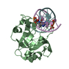
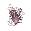
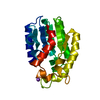
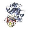
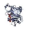
| Title | Structure of the human RAB39B in complex with GMPPNP | ||||||
 Components Components | Ras-related protein Rab-39B | ||||||
 Keywords Keywords | STRUCTURAL GENOMICS / GTPase / Ras-related protein Rab-39B / GMPPNP / Structural Genomics Consortium / SGC | ||||||
| Function / homology |  Function and homology information Function and homology informationRab protein signal transduction / RAB geranylgeranylation / myosin V binding / RAB GEFs exchange GTP for GDP on RABs / vesicle-mediated transport / cytoplasmic vesicle membrane / regulation of autophagy / synapse organization / autophagy / protein transport ...Rab protein signal transduction / RAB geranylgeranylation / myosin V binding / RAB GEFs exchange GTP for GDP on RABs / vesicle-mediated transport / cytoplasmic vesicle membrane / regulation of autophagy / synapse organization / autophagy / protein transport / vesicle / neuron projection / GTPase activity / GTP binding / Golgi apparatus / plasma membrane Similarity search - Function | ||||||
| Biological species |  Homo sapiens (human) Homo sapiens (human) | ||||||
| Method |  X-RAY DIFFRACTION / SYNCHROTRON / MOLECULAR REPLACEMENT / Resolution: 1.7 Å X-RAY DIFFRACTION / SYNCHROTRON / MOLECULAR REPLACEMENT / Resolution: 1.7 Å | ||||||
 Authors Authors | Diaz-Saez, L. / Jung, S. / von Delft, F. / Arrowsmith, C.H. / Edwards, A. / Bountra, C. / Huber, K. / Structural Genomics Consortium (SGC) | ||||||
| Funding support |  United Kingdom, 1items United Kingdom, 1items
| ||||||
 Citation Citation | Journal: To Be Published Title: Structure of the human RAB39B in complex with GMPPNP Authors: Diaz-Saez, L. / Jung, S. / Huber, K. / von Delft, F. / Arrowsmith, C.H. / Edwards, A. / Bountra, C. | ||||||
| History |
|
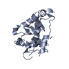
- Structure visualization
| Structure viewer | Molecule: MolmilJmol/JSmol |
|---|
- Downloads & links
-Download
| PDBx/mmCIF format | 6s5f.cif.gz | 60.8 KB | Display | PDBx/mmCIF format |
|---|---|---|---|---|
| PDB format | pdb6s5f.ent.gz | 41.6 KB | Display | PDB format |
| PDBx/mmJSON format | 6s5f.json.gz | Tree view | PDBx/mmJSON format | |
| Others |  Other downloads Other downloads |
-Validation report
| Arichive directory | https://data.pdbj.org/pub/pdb/validation_reports/s5/6s5fftp://data.pdbj.org/pub/pdb/validation_reports/s5/6s5f | HTTPS FTP |
|---|
-Related structure data
| Related structure data | 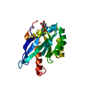 2a5jS S: Starting model for refinement |
|---|---|
| Similar structure data | |
| Other databases |


-Links
 PDBj
PDBj


- Assembly
| Deposited unit | 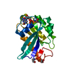
| ||||||||
|---|---|---|---|---|---|---|---|---|---|
| 1 |
| ||||||||
| Unit cell |
|
-Components
-Protein , 1 types, 1 molecules A
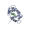
| #1: Protein | Mass: 23981.273 Da / Num. of mol.: 1 Source method: isolated from a genetically manipulated source Source: (gene. exp.) Homo sapiens (human) / Gene: RAB39B / Production host:  |
|---|
-Non-polymers , 5 types, 111 molecules 


| #2: Chemical | ChemComp-GLY /  Type: peptide linking / Mass: 75.067 Da / Num. of mol.: 1 / Source method: obtained synthetically / Formula: C2H5NO2 Type: peptide linking / Mass: 75.067 Da / Num. of mol.: 1 / Source method: obtained synthetically / Formula: C2H5NO2 |
|---|---|
| #3: Chemical | ChemComp-GNP /  Mass: 522.196 Da / Num. of mol.: 1 / Source method: obtained synthetically / Formula: C10H17N6O13P3 / Feature type: SUBJECT OF INVESTIGATION Mass: 522.196 Da / Num. of mol.: 1 / Source method: obtained synthetically / Formula: C10H17N6O13P3 / Feature type: SUBJECT OF INVESTIGATIONComment: GppNHp, GMPPNP, energy-carrying molecule analogue*YM |
| #4: Chemical | ChemComp-EDO /  Mass: 62.068 Da / Num. of mol.: 1 / Source method: obtained synthetically / Formula: C2H6O2 Mass: 62.068 Da / Num. of mol.: 1 / Source method: obtained synthetically / Formula: C2H6O2 |
| #5: Chemical | ChemComp-MG /  Mass: 24.305 Da / Num. of mol.: 1 / Source method: obtained synthetically / Formula: Mg / Feature type: SUBJECT OF INVESTIGATION Mass: 24.305 Da / Num. of mol.: 1 / Source method: obtained synthetically / Formula: Mg / Feature type: SUBJECT OF INVESTIGATION |
| #6: Water | ChemComp-HOH / Mass: 18.015 Da / Num. of mol.: 107 / Source method: isolated from a natural source / Formula: H2O |
-Details
| Has ligand of interest | Y |
|---|
-Experimental details
-Experiment
| Experiment | Method: X-RAY DIFFRACTION / Number of used crystals: 1 |
|---|
- Sample preparation
| Crystal | Density Matthews: 1.98 Å3/Da / Density % sol: 37.97 % |
|---|---|
| Crystal grow | Temperature: 293 K / Method: vapor diffusion, sitting drop / pH: 7.1 / Details: 0.1 M HEPES pH 7.1, 19 % PEG3350 |
-Data collection
| Diffraction | Mean temperature: 80 K / Serial crystal experiment: N | ||||||||||||||||||||||||||||||||||||
|---|---|---|---|---|---|---|---|---|---|---|---|---|---|---|---|---|---|---|---|---|---|---|---|---|---|---|---|---|---|---|---|---|---|---|---|---|---|
| Diffraction source | Source: SYNCHROTRON / Site: Diamond / Beamline: I03 / Wavelength: 0.91587 Å | ||||||||||||||||||||||||||||||||||||
| Detector | Type: DECTRIS EIGER2 X 16M / Detector: PIXEL / Date: May 17, 2019 | ||||||||||||||||||||||||||||||||||||
| Radiation | Protocol: SINGLE WAVELENGTH / Monochromatic (M) / Laue (L): M / Scattering type: x-ray | ||||||||||||||||||||||||||||||||||||
| Radiation wavelength | Wavelength: 0.91587 Å / Relative weight: 1 | ||||||||||||||||||||||||||||||||||||
| Reflection | Resolution: 1.7→50.28 Å / Num. obs: 20997 / % possible obs: 99.9 % / Redundancy: 6.4 % / CC1/2: 0.997 / Rmerge(I) obs: 0.105 / Rpim(I) all: 0.045 / Rrim(I) all: 0.115 / Net I/σ(I): 8.8 / Num. measured all: 134548 / Scaling rejects: 294 | ||||||||||||||||||||||||||||||||||||
| Reflection shell | Diffraction-ID: 1
|
- Processing
| Software |
| ||||||||||||||||||||||||||||||||||||||||||||||||||||||||||||
|---|---|---|---|---|---|---|---|---|---|---|---|---|---|---|---|---|---|---|---|---|---|---|---|---|---|---|---|---|---|---|---|---|---|---|---|---|---|---|---|---|---|---|---|---|---|---|---|---|---|---|---|---|---|---|---|---|---|---|---|---|---|
| Refinement | Method to determine structure: MOLECULAR REPLACEMENT Starting model: 2A5J Resolution: 1.7→50.28 Å / Cor.coef. Fo:Fc: 0.96 / Cor.coef. Fo:Fc free: 0.937 / SU B: 2.777 / SU ML: 0.089 / Cross valid method: THROUGHOUT / σ(F): 0 / ESU R: 0.117 / ESU R Free: 0.117 Details: HYDROGENS HAVE BEEN ADDED IN THE RIDING POSITIONS U VALUES : REFINED INDIVIDUALLY
| ||||||||||||||||||||||||||||||||||||||||||||||||||||||||||||
| Solvent computation | Ion probe radii: 0.8 Å / Shrinkage radii: 0.8 Å / VDW probe radii: 1.2 Å | ||||||||||||||||||||||||||||||||||||||||||||||||||||||||||||
| Displacement parameters | Biso max: 68.26 Å2 / Biso mean: 23.516 Å2 / Biso min: 13.68 Å2
| ||||||||||||||||||||||||||||||||||||||||||||||||||||||||||||
| Refinement step | Cycle: final / Resolution: 1.7→50.28 Å
| ||||||||||||||||||||||||||||||||||||||||||||||||||||||||||||
| Refine LS restraints |
| ||||||||||||||||||||||||||||||||||||||||||||||||||||||||||||
| LS refinement shell | Resolution: 1.7→1.744 Å / Rfactor Rfree error: 0 / Total num. of bins used: 20
|
