 Movie
Movie Controller

[English] 日本語
 Yorodumi
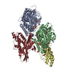
- PDB-6sjc: Structure of T. thermophilus AspRS in Complex with 5'-O-(N-(L-asp... -
+ Open data

- Basic information
| Entry | Database: PDB / ID: 6sjc | ||||||||||||
|---|---|---|---|---|---|---|---|---|---|---|---|---|---|
| Title | Structure of T. thermophilus AspRS in Complex with 5'-O-(N-(L-aspartyl)-sulfamoyl)adenosine | ||||||||||||
 Components Components | Aspartate--tRNA(Asp) ligase | ||||||||||||
 Keywords Keywords | LIGASE / protein-inhibitor complex / tRNA aminoacylation | ||||||||||||
| Function / homology |  Function and homology information Function and homology informationaspartate-tRNA ligase / aspartate-tRNA ligase activity / aspartyl-tRNA aminoacylation / nucleic acid binding / ATP binding / cytoplasm Similarity search - Function | ||||||||||||
| Biological species |   Thermus thermophilus (bacteria) Thermus thermophilus (bacteria) | ||||||||||||
| Method |  X-RAY DIFFRACTION / SYNCHROTRON / MOLECULAR REPLACEMENT / Resolution: 2.23 Å X-RAY DIFFRACTION / SYNCHROTRON / MOLECULAR REPLACEMENT / Resolution: 2.23 Å | ||||||||||||
 Authors Authors | De Graef, S. / Pang, L. / Strelkov, S.V. / Weeks, S.D. | ||||||||||||
| Funding support |  Belgium, 3items Belgium, 3items
| ||||||||||||
 Citation Citation | Journal: Acs Chem.Biol. / Year: 2020 Title: Structural Insights into the Binding of Natural Pyrimidine-Based Inhibitors of Class II Aminoacyl-tRNA Synthetases. Authors: Pang, L. / Nautiyal, M. / De Graef, S. / Gadakh, B. / Zorzini, V. / Economou, A. / Strelkov, S.V. / Van Aerschot, A. / Weeks, S.D. | ||||||||||||
| History |
|
- Structure visualization
| Structure viewer | Molecule: MolmilJmol/JSmol |
|---|
- Downloads & links
-Download
| PDBx/mmCIF format | 6sjc.cif.gz | 445.6 KB | Display | PDBx/mmCIF format |
|---|---|---|---|---|
| PDB format | pdb6sjc.ent.gz | 364.1 KB | Display | PDB format |
| PDBx/mmJSON format | 6sjc.json.gz | Tree view | PDBx/mmJSON format | |
| Others |  Other downloads Other downloads |
-Validation report
| Arichive directory | https://data.pdbj.org/pub/pdb/validation_reports/sj/6sjcftp://data.pdbj.org/pub/pdb/validation_reports/sj/6sjc | HTTPS FTP |
|---|
-Related structure data
| Related structure data | 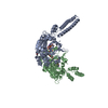 6hdzC  6he1C  6he3C 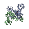 6hhvC 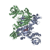 6hhwC  6hhxC 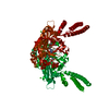 6hhyC 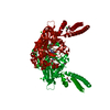 6hhzC  6hi0C  6rltC 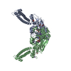 6rluC 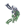 6rlvC  6s30C  1lowS C: citing same article ( S: Starting model for refinement |
|---|---|
| Similar structure data |


-Links
 PDBj
PDBj

- Assembly
| Deposited unit | 
| ||||||||
|---|---|---|---|---|---|---|---|---|---|
| 1 |
| ||||||||
| Unit cell |
|
-Components
| #1: Protein | Mass: 66181.828 Da / Num. of mol.: 2 / Fragment: aspartyl-tRNA synthetase Source method: isolated from a genetically manipulated source Source: (gene. exp.) Thermus thermophilus (bacteria) / Gene: aspS / Plasmid: pETRUK / Production host: References: UniProt: P36419, UniProt: Q5SKD2*PLUS, aspartate-tRNA ligase #2: Chemical | 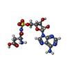  Mass: 461.407 Da / Num. of mol.: 2 / Source method: obtained synthetically / Formula: C14H19N7O9S / Feature type: SUBJECT OF INVESTIGATION Mass: 461.407 Da / Num. of mol.: 2 / Source method: obtained synthetically / Formula: C14H19N7O9S / Feature type: SUBJECT OF INVESTIGATION#3: Water | ChemComp-HOH / |  Mass: 18.015 Da / Num. of mol.: 193 / Source method: isolated from a natural source / Formula: H2O Mass: 18.015 Da / Num. of mol.: 193 / Source method: isolated from a natural source / Formula: H2OHas ligand of interest | Y | |
|---|
-Experimental details
-Experiment
| Experiment | Method: X-RAY DIFFRACTION / Number of used crystals: 1 |
|---|
- Sample preparation
| Crystal | Density Matthews: 3.01 Å3/Da / Density % sol: 59.18 % |
|---|---|
| Crystal grow | Temperature: 293 K / Method: vapor diffusion, hanging drop / pH: 7 Details: A 10 mg/ml protein solution was prepared in 10 mM TRIS pH 7.5, 100 mM NaCl, 2.5 mM DTT and 0.4% w/v low melting point agarose, maintaining the sample temperature at 315 kelvin. Crystals were ...Details: A 10 mg/ml protein solution was prepared in 10 mM TRIS pH 7.5, 100 mM NaCl, 2.5 mM DTT and 0.4% w/v low melting point agarose, maintaining the sample temperature at 315 kelvin. Crystals were grown by mixing an equal volume of the protein solution with 8-12% PEG 4000, 0.1 M Morpheus buffer system 1 (MES/imidazole) pH 7, 100 mM KCl, 20 v/v % glycerol. For soaking a 4 mM solution of compound in DMSO was used. A one third volume of the initial drop size was pipetted carefully onto the crystal containing drop. The sample was then placed back over the reservoir and incubated for approximately 2 hr. Crystals were caught in cryoloops and directly flash frozen in liquid nitrogen. |
-Data collection
| Diffraction | Mean temperature: 100 K / Serial crystal experiment: N | ||||||||||||||||||||||||||||||
|---|---|---|---|---|---|---|---|---|---|---|---|---|---|---|---|---|---|---|---|---|---|---|---|---|---|---|---|---|---|---|---|
| Diffraction source | Source: SYNCHROTRON / Site: SOLEIL  / Beamline: PROXIMA 1 / Wavelength: 0.978565 Å / Beamline: PROXIMA 1 / Wavelength: 0.978565 Å | ||||||||||||||||||||||||||||||
| Detector | Type: DECTRIS EIGER X 16M / Detector: PIXEL / Date: Feb 15, 2019 | ||||||||||||||||||||||||||||||
| Radiation | Protocol: SINGLE WAVELENGTH / Monochromatic (M) / Laue (L): M / Scattering type: x-ray | ||||||||||||||||||||||||||||||
| Radiation wavelength | Wavelength: 0.978565 Å / Relative weight: 1 | ||||||||||||||||||||||||||||||
| Reflection | Resolution: 2.16→79.59 Å / Num. obs: 83261 / % possible obs: 99.7 % / Redundancy: 7 % / CC1/2: 0.999 / Rmerge(I) obs: 0.064 / Rpim(I) all: 0.026 / Rrim(I) all: 0.069 / Net I/σ(I): 13.4 / Num. measured all: 583963 | ||||||||||||||||||||||||||||||
| Reflection shell | Diffraction-ID: 1
|
- Processing
| Software |
| ||||||||||||||||||||||||||||||||||||||||||||||||||||||||||||||||||||||||||||||||||||||||||||||||||||||
|---|---|---|---|---|---|---|---|---|---|---|---|---|---|---|---|---|---|---|---|---|---|---|---|---|---|---|---|---|---|---|---|---|---|---|---|---|---|---|---|---|---|---|---|---|---|---|---|---|---|---|---|---|---|---|---|---|---|---|---|---|---|---|---|---|---|---|---|---|---|---|---|---|---|---|---|---|---|---|---|---|---|---|---|---|---|---|---|---|---|---|---|---|---|---|---|---|---|---|---|---|---|---|---|
| Refinement | Method to determine structure: MOLECULAR REPLACEMENT Starting model: 1LOW Resolution: 2.23→57.581 Å / SU ML: 0.3 / Cross valid method: THROUGHOUT / σ(F): 1.37 / Phase error: 26.91 / Stereochemistry target values: ML
| ||||||||||||||||||||||||||||||||||||||||||||||||||||||||||||||||||||||||||||||||||||||||||||||||||||||
| Solvent computation | Shrinkage radii: 0.9 Å / VDW probe radii: 1.11 Å / Solvent model: FLAT BULK SOLVENT MODEL | ||||||||||||||||||||||||||||||||||||||||||||||||||||||||||||||||||||||||||||||||||||||||||||||||||||||
| Displacement parameters | Biso max: 174.84 Å2 / Biso mean: 67.9309 Å2 / Biso min: 34.14 Å2 | ||||||||||||||||||||||||||||||||||||||||||||||||||||||||||||||||||||||||||||||||||||||||||||||||||||||
| Refinement step | Cycle: final / Resolution: 2.23→57.581 Å
| ||||||||||||||||||||||||||||||||||||||||||||||||||||||||||||||||||||||||||||||||||||||||||||||||||||||
| LS refinement shell | Refine-ID: X-RAY DIFFRACTION / Rfactor Rfree error: 0
| ||||||||||||||||||||||||||||||||||||||||||||||||||||||||||||||||||||||||||||||||||||||||||||||||||||||
| Refinement TLS params. | Method: refined / Refine-ID: X-RAY DIFFRACTION
| ||||||||||||||||||||||||||||||||||||||||||||||||||||||||||||||||||||||||||||||||||||||||||||||||||||||
| Refinement TLS group |
|
