 Movie
Movie Controller

[English] 日本語
 Yorodumi
- PDB-7ap4: Thermus thermophilus Aspartyl-tRNA Synthetase in Complex with Com... -
+ Open data

- Basic information
| Entry | Database: PDB / ID: 7ap4 | ||||||
|---|---|---|---|---|---|---|---|
| Title | Thermus thermophilus Aspartyl-tRNA Synthetase in Complex with Compound AspS7HMDDA | ||||||
 Components Components | Aspartate--tRNA(Asp) ligase | ||||||
 Keywords Keywords | LIGASE / protein-inhibitor complex / tRNA aminoacylation | ||||||
| Function / homology |  Function and homology information Function and homology informationaspartate-tRNA ligase / aspartate-tRNA ligase activity / aspartyl-tRNA aminoacylation / nucleic acid binding / ATP binding / cytoplasm Similarity search - Function | ||||||
| Biological species |   Thermus thermophilus (bacteria) Thermus thermophilus (bacteria) | ||||||
| Method |  X-RAY DIFFRACTION / SYNCHROTRON / MOLECULAR REPLACEMENT / Resolution: 2.15 Å X-RAY DIFFRACTION / SYNCHROTRON / MOLECULAR REPLACEMENT / Resolution: 2.15 Å | ||||||
 Authors Authors | De Graef, S. / Pang, L. / Strelkov, S.V. / Weeks, S.D. | ||||||
| Funding support |  Belgium, 1items Belgium, 1items
| ||||||
 Citation Citation | Journal: Molecules / Year: 2020 Title: Synthesis and Biological Evaluation of 1,3-Dideazapurine-Like 7-Amino-5-Hydroxymethyl-Benzimidazole Ribonucleoside Analogues as Aminoacyl-tRNA Synthetase Inhibitors. Authors: Zhang, B. / Pang, L. / Nautiyal, M. / De Graef, S. / Gadakh, B. / Lescrinier, E. / Rozenski, J. / Strelkov, S.V. / Weeks, S.D. / Van Aerschot, A. | ||||||
| History |
|
- Structure visualization
| Structure viewer | Molecule: MolmilJmol/JSmol |
|---|
- Downloads & links
-Download
| PDBx/mmCIF format | 7ap4.cif.gz | 429.9 KB | Display | PDBx/mmCIF format |
|---|---|---|---|---|
| PDB format | pdb7ap4.ent.gz | 353.5 KB | Display | PDB format |
| PDBx/mmJSON format | 7ap4.json.gz | Tree view | PDBx/mmJSON format | |
| Others |  Other downloads Other downloads |
-Validation report
| Arichive directory | https://data.pdbj.org/pub/pdb/validation_reports/ap/7ap4ftp://data.pdbj.org/pub/pdb/validation_reports/ap/7ap4 | HTTPS FTP |
|---|
-Related structure data
| Related structure data |  7ap1C 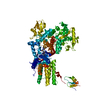 7ap2C 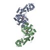 7ap3C 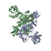 1l0wS S: Starting model for refinement C: citing same article ( |
|---|---|
| Similar structure data |


-Links
 PDBj
PDBj

- Assembly
| Deposited unit | 
| ||||||||
|---|---|---|---|---|---|---|---|---|---|
| 1 |
| ||||||||
| Unit cell |
|
-Components
| #1: Protein | Mass: 66181.828 Da / Num. of mol.: 2 Source method: isolated from a genetically manipulated source Source: (gene. exp.) Thermus thermophilus (strain HB8 / ATCC 27634 / DSM 579) (bacteria)Strain: HB8 / ATCC 27634 / DSM 579 / Gene: aspS1, TTHA0711 / Plasmid: pETRUK / Production host: #2: Chemical |   Mass: 489.457 Da / Num. of mol.: 2 / Source method: obtained synthetically / Formula: C17H23N5O10S / Feature type: SUBJECT OF INVESTIGATION Mass: 489.457 Da / Num. of mol.: 2 / Source method: obtained synthetically / Formula: C17H23N5O10S / Feature type: SUBJECT OF INVESTIGATION#3: Water | ChemComp-HOH / |  Mass: 18.015 Da / Num. of mol.: 297 / Source method: isolated from a natural source / Formula: H2O Mass: 18.015 Da / Num. of mol.: 297 / Source method: isolated from a natural source / Formula: H2OHas ligand of interest | Y | |
|---|
-Experimental details
-Experiment
| Experiment | Method: X-RAY DIFFRACTION / Number of used crystals: 1 |
|---|
- Sample preparation
| Crystal | Density Matthews: 3.01 Å3/Da / Density % sol: 59.14 % |
|---|---|
| Crystal grow | Temperature: 293 K / Method: vapor diffusion / pH: 7 Details: A 10 mg/ml protein solution was prepared in 10 mM TRIS pH 7.5, 100 mM NaCl, 2.5 mM DTT and 0.4% w/v low melting point agarose, maintaining the sample temperature at 315 kelvin. Crystals were ...Details: A 10 mg/ml protein solution was prepared in 10 mM TRIS pH 7.5, 100 mM NaCl, 2.5 mM DTT and 0.4% w/v low melting point agarose, maintaining the sample temperature at 315 kelvin. Crystals were grown by mixing an equal volume of the protein solution with 8-12% PEG 4000, 0.1 M Morpheus buffer system 1 (MES/imidazole) pH 7, 100 mM KCl, 20 v/v % glycerol. For soaking a 4 mM solution of compound in DMSO was used. A one third volume of the initial drop size was pipetted carefully onto the crystal containing drop. The sample was then placed back over the reservoir and incubated for approximately 2 hr. Crystals were caught in cryoloops and directly flash frozen in liquid nitrogen. |
-Data collection
| Diffraction | Mean temperature: 100 K / Serial crystal experiment: N | ||||||||||||||||||||||||||||||
|---|---|---|---|---|---|---|---|---|---|---|---|---|---|---|---|---|---|---|---|---|---|---|---|---|---|---|---|---|---|---|---|
| Diffraction source | Source: SYNCHROTRON / Site: ESRF  / Beamline: ID29 / Wavelength: 0.96863 Å / Beamline: ID29 / Wavelength: 0.96863 Å | ||||||||||||||||||||||||||||||
| Detector | Type: DECTRIS PILATUS 6M / Detector: PIXEL / Date: Feb 16, 2018 | ||||||||||||||||||||||||||||||
| Radiation | Protocol: SINGLE WAVELENGTH / Monochromatic (M) / Laue (L): M / Scattering type: x-ray | ||||||||||||||||||||||||||||||
| Radiation wavelength | Wavelength: 0.96863 Å / Relative weight: 1 | ||||||||||||||||||||||||||||||
| Reflection | Resolution: 2.06→79.53 Å / Num. obs: 94888 / % possible obs: 99 % / Redundancy: 3.8 % / Biso Wilson estimate: 47.89 Å2 / CC1/2: 0.997 / Rmerge(I) obs: 0.071 / Rpim(I) all: 0.041 / Rrim(I) all: 0.082 / Net I/σ(I): 9.5 / Num. measured all: 356281 / Scaling rejects: 18 | ||||||||||||||||||||||||||||||
| Reflection shell | Diffraction-ID: 1
|
- Processing
| Software |
| ||||||||||||||||||||||||||||||||||||||||||||||||||||||||||||||||||||||||||||||||||||||||||||||||
|---|---|---|---|---|---|---|---|---|---|---|---|---|---|---|---|---|---|---|---|---|---|---|---|---|---|---|---|---|---|---|---|---|---|---|---|---|---|---|---|---|---|---|---|---|---|---|---|---|---|---|---|---|---|---|---|---|---|---|---|---|---|---|---|---|---|---|---|---|---|---|---|---|---|---|---|---|---|---|---|---|---|---|---|---|---|---|---|---|---|---|---|---|---|---|---|---|---|
| Refinement | Method to determine structure: MOLECULAR REPLACEMENT Starting model: 1L0W Resolution: 2.15→79.526 Å / SU ML: 0.29 / Cross valid method: THROUGHOUT / σ(F): 0.31 / Phase error: 26.6 / Stereochemistry target values: ML
| ||||||||||||||||||||||||||||||||||||||||||||||||||||||||||||||||||||||||||||||||||||||||||||||||
| Solvent computation | Shrinkage radii: 0.9 Å / VDW probe radii: 1.11 Å / Solvent model: FLAT BULK SOLVENT MODEL | ||||||||||||||||||||||||||||||||||||||||||||||||||||||||||||||||||||||||||||||||||||||||||||||||
| Displacement parameters | Biso max: 553.7 Å2 / Biso mean: 70.5674 Å2 / Biso min: 30.07 Å2 | ||||||||||||||||||||||||||||||||||||||||||||||||||||||||||||||||||||||||||||||||||||||||||||||||
| Refinement step | Cycle: final / Resolution: 2.15→79.526 Å
| ||||||||||||||||||||||||||||||||||||||||||||||||||||||||||||||||||||||||||||||||||||||||||||||||
| LS refinement shell | Refine-ID: X-RAY DIFFRACTION / Rfactor Rfree error: 0
|
