 Movie
Movie Controller

[English] 日本語
 Yorodumi
- PDB-1low: X-ray structure of the H40A mutant of Ribonuclease T1 complexed w... -
+ Open data

- Basic information
| Entry | Database: PDB / ID: 1low | ||||||
|---|---|---|---|---|---|---|---|
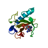
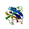
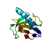
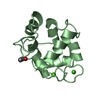
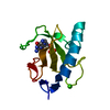
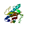
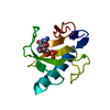
| Title | X-ray structure of the H40A mutant of Ribonuclease T1 complexed with 3'-guanosine monophosphate | ||||||
 Components Components | Guanyl-specific ribonuclease T1 | ||||||
 Keywords Keywords | HYDROLASE / RNase / catalytic dyad / nucleophile activation / ab initio calculations | ||||||
| Function / homology |  Function and homology information Function and homology informationhyphal tip / ribonuclease T1 / ribonuclease T1 activity / cell septum / hydrolase activity / RNA binding Similarity search - Function | ||||||
| Biological species |  | ||||||
| Method |  X-RAY DIFFRACTION / MOLECULAR REPLACEMENT / Resolution: 1.9 Å X-RAY DIFFRACTION / MOLECULAR REPLACEMENT / Resolution: 1.9 Å | ||||||
 Authors Authors | Mignon, P. / Steyaert, J. / Loris, R. / Geerlings, P. / Loverix, S. | ||||||
 Citation Citation | Journal: J.Biol.Chem. / Year: 2002 Title: A nucleophile activation dyad in ribonucleases. A combined X-ray crystallographic/ab initio quantum chemical study Authors: Mignon, P. / Steyaert, J. / Loris, R. / Geerlings, P. / Loverix, S. | ||||||
| History |
| ||||||
| Remark 999 | The isozyme of RNase T1 used here contains a lysine at position 25. |
- Structure visualization
| Structure viewer | Molecule: MolmilJmol/JSmol |
|---|
- Downloads & links
-Download
| PDBx/mmCIF format | 1low.cif.gz | 33.8 KB | Display | PDBx/mmCIF format |
|---|---|---|---|---|
| PDB format | pdb1low.ent.gz | 21.7 KB | Display | PDB format |
| PDBx/mmJSON format | 1low.json.gz | Tree view | PDBx/mmJSON format | |
| Others |  Other downloads Other downloads |
-Validation report
| Arichive directory | https://data.pdbj.org/pub/pdb/validation_reports/lo/1lowftp://data.pdbj.org/pub/pdb/validation_reports/lo/1low | HTTPS FTP |
|---|
-Related structure data
| Related structure data | 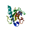 1lovC 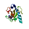 1loyC 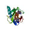 1rgaS S: Starting model for refinement C: citing same article ( |
|---|---|
| Similar structure data |


-Links
 PDBj
PDBj- Assembly
| Deposited unit | 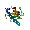
| ||||||||
|---|---|---|---|---|---|---|---|---|---|
| 1 |
| ||||||||
| Unit cell |
|
-Components
| #1: Protein | Mass: 11027.625 Da / Num. of mol.: 1 / Mutation: H40A Source method: isolated from a genetically manipulated source Source: (gene. exp.)  |
|---|---|
| #2: Chemical | ChemComp-CA /   Mass: 40.078 Da / Num. of mol.: 1 / Source method: obtained synthetically / Formula: Ca Mass: 40.078 Da / Num. of mol.: 1 / Source method: obtained synthetically / Formula: Ca |
| #3: Chemical | ChemComp-3GP /   Mass: 363.221 Da / Num. of mol.: 1 / Source method: obtained synthetically / Formula: C10H14N5O8P Mass: 363.221 Da / Num. of mol.: 1 / Source method: obtained synthetically / Formula: C10H14N5O8P |
| #4: Water | ChemComp-HOH /  Mass: 18.015 Da / Num. of mol.: 35 / Source method: isolated from a natural source / Formula: H2O Mass: 18.015 Da / Num. of mol.: 35 / Source method: isolated from a natural source / Formula: H2O |
| Has protein modification | Y |
-Experimental details
-Experiment
| Experiment | Method: X-RAY DIFFRACTION / Number of used crystals: 1 |
|---|
- Sample preparation
| Crystal | Density Matthews: 2.09 Å3/Da / Density % sol: 41.08 % | ||||||||||||||||||||||||||||||||||||||||||
|---|---|---|---|---|---|---|---|---|---|---|---|---|---|---|---|---|---|---|---|---|---|---|---|---|---|---|---|---|---|---|---|---|---|---|---|---|---|---|---|---|---|---|---|
| Crystal grow | Temperature: 293 K / Method: vapor diffusion, hanging drop / pH: 4.2 Details: MPD,CaCl2,sodium acetate, pH 4.2, VAPOR DIFFUSION, HANGING DROP, temperature 293K | ||||||||||||||||||||||||||||||||||||||||||
| Crystal grow | *PLUS Temperature: 20 ℃ / pH: 7.2 | ||||||||||||||||||||||||||||||||||||||||||
| Components of the solutions | *PLUS
|
-Data collection
| Diffraction | Mean temperature: 293 K |
|---|---|
| Diffraction source | Source: ROTATING ANODE / Type: RIGAKU RU200 / Wavelength: 1.5418 Å |
| Detector | Type: MARRESEARCH / Detector: IMAGE PLATE / Date: Mar 21, 2001 |
| Radiation | Monochromator: Graphite / Protocol: SINGLE WAVELENGTH / Monochromatic (M) / Laue (L): M / Scattering type: x-ray |
| Radiation wavelength | Wavelength: 1.5418 Å / Relative weight: 1 |
| Reflection | Resolution: 1.9→15 Å / Num. all: 7614 / Num. obs: 7614 / % possible obs: 99.6 % / Observed criterion σ(I): -3 / Redundancy: 7.5 % / Biso Wilson estimate: 25.2 Å2 / Rmerge(I) obs: 0.102 / Net I/σ(I): 14.94 |
| Reflection shell | Resolution: 1.9→1.97 Å / Rmerge(I) obs: 0.785 / Mean I/σ(I) obs: 3.7 / % possible all: 98.9 |
| Reflection | *PLUS Highest resolution: 1.9 Å / Num. obs: 7613 / % possible obs: 99.31 % |
- Processing
| Software |
| ||||||||||||||||||||
|---|---|---|---|---|---|---|---|---|---|---|---|---|---|---|---|---|---|---|---|---|---|
| Refinement | Method to determine structure: MOLECULAR REPLACEMENT Starting model: PDB ENTRY 1RGA Resolution: 1.9→22.8 Å / Cross valid method: THROUGHOUT / σ(F): 0.001 / Stereochemistry target values: Engh & Huber
| ||||||||||||||||||||
| Refinement step | Cycle: LAST / Resolution: 1.9→22.8 Å
| ||||||||||||||||||||
| Refine LS restraints |
| ||||||||||||||||||||
| Refinement | *PLUS % reflection Rfree: 10 % / Rfactor Rfree: 0.23 / Rfactor Rwork: 0.205 | ||||||||||||||||||||
| Solvent computation | *PLUS | ||||||||||||||||||||
| Displacement parameters | *PLUS | ||||||||||||||||||||
| Refine LS restraints | *PLUS
|
