 Movie
Movie Controller

[English] 日本語
 Yorodumi
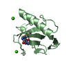
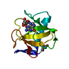
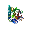
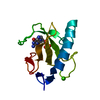
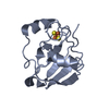
- PDB-1fus: CRYSTAL STRUCTURES OF RIBONUCLEASE F1 OF FUSARIUM MONILIFORME IN ... -
+ Open data

- Basic information
| Entry | Database: PDB / ID: 1fus | |||||||||
|---|---|---|---|---|---|---|---|---|---|---|
| Title | CRYSTAL STRUCTURES OF RIBONUCLEASE F1 OF FUSARIUM MONILIFORME IN ITS FREE FORM AND IN COMPLEX WITH 2'GMP | |||||||||
 Components Components | RIBONUCLEASE F1 | |||||||||
 Keywords Keywords | HYDROLASE(ENDORIBONUCLEASE) | |||||||||
| Function / homology |  Function and homology information Function and homology informationribonuclease T1 / ribonuclease T1 activity / hydrolase activity / RNA binding Similarity search - Function | |||||||||
| Biological species |  Gibberella fujikuroi (fungus) Gibberella fujikuroi (fungus) | |||||||||
| Method |  X-RAY DIFFRACTION / Resolution: 1.3 Å X-RAY DIFFRACTION / Resolution: 1.3 Å | |||||||||
 Authors Authors | Katayanagi, K. / Vassylyev, D.G. / Ishikawa, K. / Morikawa, K. | |||||||||
 Citation Citation | Journal: J.Mol.Biol. / Year: 1993 Title: Crystal structures of ribonuclease F1 of Fusarium moniliforme in its free form and in complex with 2'GMP. Authors: Vassylyev, D.G. / Katayanagi, K. / Ishikawa, K. / Tsujimoto-Hirano, M. / Danno, M. / Pahler, A. / Matsumoto, O. / Matsushima, M. / Yoshida, H. / Morikawa, K. | |||||||||
| History |
|
- Structure visualization
| Structure viewer | Molecule: MolmilJmol/JSmol |
|---|
- Downloads & links
-Download
| PDBx/mmCIF format | 1fus.cif.gz | 33.2 KB | Display | PDBx/mmCIF format |
|---|---|---|---|---|
| PDB format | pdb1fus.ent.gz | 21.8 KB | Display | PDB format |
| PDBx/mmJSON format | 1fus.json.gz | Tree view | PDBx/mmJSON format | |
| Others |  Other downloads Other downloads |
-Validation report
| Arichive directory | https://data.pdbj.org/pub/pdb/validation_reports/fu/1fusftp://data.pdbj.org/pub/pdb/validation_reports/fu/1fus | HTTPS FTP |
|---|
-Related structure data


-Links
 PDBj
PDBj- Assembly
| Deposited unit | 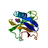
| ||||||||
|---|---|---|---|---|---|---|---|---|---|
| 1 |
| ||||||||
| Unit cell |
| ||||||||
| Atom site foot note | 1: RESIDUES 39 AND 55 ARE CIS PROLINES. |
-Components
| #1: Protein | Mass: 10989.544 Da / Num. of mol.: 1 Source method: isolated from a genetically manipulated source Source: (gene. exp.) Gibberella fujikuroi (fungus) / References: UniProt: P10282, EC: 3.1.27.3 |
|---|---|
| #2: Water | ChemComp-HOH /  Mass: 18.015 Da / Num. of mol.: 107 / Source method: isolated from a natural source / Formula: H2O Mass: 18.015 Da / Num. of mol.: 107 / Source method: isolated from a natural source / Formula: H2O |
| Has protein modification | Y |
| Sequence details | SEQUENCE ADVISORY NOTICE: DIFFERENCE BETWEEN SWISS-PROT AND PDB SEQUENCE. SWISS-PROT ENTRY NAME: ...SEQUENCE ADVISORY NOTICE: DIFFERENCE |
-Experimental details
-Experiment
| Experiment | Method: X-RAY DIFFRACTION |
|---|
- Sample preparation
| Crystal | Density Matthews: 1.89 Å3/Da / Density % sol: 34.78 % | ||||||||||||||||||||
|---|---|---|---|---|---|---|---|---|---|---|---|---|---|---|---|---|---|---|---|---|---|
| Crystal grow | *PLUS Temperature: 20 ℃ / pH: 3.5 / Method: vapor diffusion, hanging drop | ||||||||||||||||||||
| Components of the solutions | *PLUS
|
-Data collection
| Radiation | Scattering type: x-ray |
|---|---|
| Radiation wavelength | Relative weight: 1 |
| Reflection | *PLUS Num. obs: 13257 |
- Processing
| Software | Name: PROLSQ / Classification: refinement | ||||||||||||||||||||||||||||||||||||||||||||||||||||||||||||||||||||||||||||||||||||
|---|---|---|---|---|---|---|---|---|---|---|---|---|---|---|---|---|---|---|---|---|---|---|---|---|---|---|---|---|---|---|---|---|---|---|---|---|---|---|---|---|---|---|---|---|---|---|---|---|---|---|---|---|---|---|---|---|---|---|---|---|---|---|---|---|---|---|---|---|---|---|---|---|---|---|---|---|---|---|---|---|---|---|---|---|---|
| Refinement | Resolution: 1.3→8 Å / Rfactor obs: 0.187 / σ(F): 1 | ||||||||||||||||||||||||||||||||||||||||||||||||||||||||||||||||||||||||||||||||||||
| Refinement step | Cycle: LAST / Resolution: 1.3→8 Å
| ||||||||||||||||||||||||||||||||||||||||||||||||||||||||||||||||||||||||||||||||||||
| Refine LS restraints |
| ||||||||||||||||||||||||||||||||||||||||||||||||||||||||||||||||||||||||||||||||||||
| Refinement | *PLUS Highest resolution: 1.3 Å / Lowest resolution: 8 Å / σ(F): 1 / Rfactor obs: 0.187 | ||||||||||||||||||||||||||||||||||||||||||||||||||||||||||||||||||||||||||||||||||||
| Solvent computation | *PLUS | ||||||||||||||||||||||||||||||||||||||||||||||||||||||||||||||||||||||||||||||||||||
| Displacement parameters | *PLUS |
