 Movie
Movie Controller


+ Open data

- Basic information
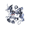
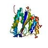
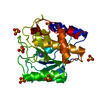
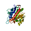
| Entry | Database: PDB / ID: 6s8r | ||||||
|---|---|---|---|---|---|---|---|
| Title | D. melanogaster RNA helicase Me31B in complex with GIGYF | ||||||
 Components Components |
| ||||||
 Keywords Keywords | RNA BINDING PROTEIN / Translation / Translational control / mRNA decay / miRNA | ||||||
| Function / homology |  Function and homology information Function and homology informationpositive regulation of post-transcriptional gene silencing / sensory dendrite / regulation of olfactory learning / mRNA decay by 5' to 3' exoribonuclease / regulation of neuron projection arborization / pole cell formation / follicle cell of egg chamber development / deadenylation-independent decapping of nuclear-transcribed mRNA / neuronal ribonucleoprotein granule / positive regulation of mRNA catabolic process ...positive regulation of post-transcriptional gene silencing / sensory dendrite / regulation of olfactory learning / mRNA decay by 5' to 3' exoribonuclease / regulation of neuron projection arborization / pole cell formation / follicle cell of egg chamber development / deadenylation-independent decapping of nuclear-transcribed mRNA / neuronal ribonucleoprotein granule / positive regulation of mRNA catabolic process / deadenylation-dependent decapping of nuclear-transcribed mRNA / eukaryotic translation initiation factor 4F complex / miRNA-mediated post-transcriptional gene silencing / regulation of defense response to virus / habituation / miRNA-mediated gene silencing by inhibition of translation / P granule / P-body assembly / muscle cell cellular homeostasis / negative regulation of cytoplasmic translation / mRNA regulatory element binding translation repressor activity / stress granule assembly / regulation of autophagy / P-body / neuron cellular homeostasis / cytoplasmic stress granule / cytoplasmic ribonucleoprotein granule / RNA helicase activity / negative regulation of translation / postsynapse / RNA helicase / mRNA binding / neuronal cell body / endoplasmic reticulum / ATP hydrolysis activity / RNA binding / ATP binding / cytoplasm / cytosol Similarity search - Function | ||||||
| Biological species |  | ||||||
| Method |  X-RAY DIFFRACTION / SYNCHROTRON / MOLECULAR REPLACEMENT / Resolution: 2.41 Å X-RAY DIFFRACTION / SYNCHROTRON / MOLECULAR REPLACEMENT / Resolution: 2.41 Å | ||||||
 Authors Authors | Peter, D. / Valkov, E. | ||||||
 Citation Citation | Journal: Genes Dev. / Year: 2019 Title: Molecular basis for GIGYF-Me31B complex assembly in 4EHP-mediated translational repression. Authors: Peter, D. / Ruscica, V. / Bawankar, P. / Weber, R. / Helms, S. / Valkov, E. / Igreja, C. / Izaurralde, E. | ||||||
| History |
|
- Structure visualization
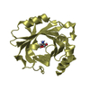
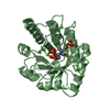
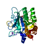
| Structure viewer | Molecule: MolmilJmol/JSmol |
|---|
- Downloads & links
-Download
| PDBx/mmCIF format | 6s8r.cif.gz | 65.3 KB | Display | PDBx/mmCIF format |
|---|---|---|---|---|
| PDB format | pdb6s8r.ent.gz | 38.3 KB | Display | PDB format |
| PDBx/mmJSON format | 6s8r.json.gz | Tree view | PDBx/mmJSON format | |
| Others |  Other downloads Other downloads |
-Validation report
| Arichive directory | https://data.pdbj.org/pub/pdb/validation_reports/s8/6s8rftp://data.pdbj.org/pub/pdb/validation_reports/s8/6s8r | HTTPS FTP |
|---|
-Related structure data
| Related structure data | 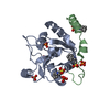 6s8sC 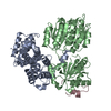 5anrS S: Starting model for refinement C: citing same article ( |
|---|---|
| Similar structure data |


-Links
 PDBj
PDBj- Assembly
| Deposited unit | 
| ||||||||||||
|---|---|---|---|---|---|---|---|---|---|---|---|---|---|
| 1 |
| ||||||||||||
| Unit cell |
|
-Components
| #1: Protein | Mass: 20004.273 Da / Num. of mol.: 1 Source method: isolated from a genetically manipulated source Source: (gene. exp.)  | ||||||
|---|---|---|---|---|---|---|---|
| #2: Protein/peptide | Mass: 2847.999 Da / Num. of mol.: 1 Source method: isolated from a genetically manipulated source Source: (gene. exp.) | ||||||
| #3: Chemical | ChemComp-ACT /   Mass: 59.044 Da / Num. of mol.: 4 / Source method: obtained synthetically / Formula: C2H3O2 Mass: 59.044 Da / Num. of mol.: 4 / Source method: obtained synthetically / Formula: C2H3O2#4: Water | ChemComp-HOH / |  Mass: 18.015 Da / Num. of mol.: 35 / Source method: isolated from a natural source / Formula: H2O Mass: 18.015 Da / Num. of mol.: 35 / Source method: isolated from a natural source / Formula: H2OHas ligand of interest | N | Has protein modification | Y | |
-Experimental details
-Experiment
| Experiment | Method: X-RAY DIFFRACTION / Number of used crystals: 1 |
|---|
- Sample preparation
| Crystal | Density Matthews: 2.15 Å3/Da / Density % sol: 42.84 % |
|---|---|
| Crystal grow | Temperature: 291 K / Method: vapor diffusion, hanging drop Details: 0.1 M sodium acetate (pH 5.0), 0.15 M ammonium chloride, 16% (w/v) PEG 6000 |
-Data collection
| Diffraction | Mean temperature: 100 K / Serial crystal experiment: N |
|---|---|
| Diffraction source | Source: SYNCHROTRON / Site: SLS  / Beamline: X10SA / Wavelength: 0.9996 Å / Beamline: X10SA / Wavelength: 0.9996 Å |
| Detector | Type: DECTRIS PILATUS 6M / Detector: PIXEL / Date: Aug 30, 2016 |
| Radiation | Protocol: SINGLE WAVELENGTH / Monochromatic (M) / Laue (L): M / Scattering type: x-ray |
| Radiation wavelength | Wavelength: 0.9996 Å / Relative weight: 1 |
| Reflection | Resolution: 2.4→42 Å / Num. obs: 7712 / % possible obs: 95.8 % / Redundancy: 10.4 % / Biso Wilson estimate: 27.21 Å2 / Rsym value: 0.188 / Net I/σ(I): 8.91 |
| Reflection shell | Resolution: 2.4→2.46 Å / Redundancy: 9.7 % / Num. unique obs: 522 / Rsym value: 1.06 / % possible all: 88.5 |
- Processing
| Software |
| ||||||||||||||||||||||||
|---|---|---|---|---|---|---|---|---|---|---|---|---|---|---|---|---|---|---|---|---|---|---|---|---|---|
| Refinement | Method to determine structure: MOLECULAR REPLACEMENT Starting model: 5ANR Resolution: 2.41→39.7 Å / SU ML: 0.2761 / Cross valid method: FREE R-VALUE / σ(F): 1.37 / Phase error: 26.1878
| ||||||||||||||||||||||||
| Solvent computation | Shrinkage radii: 0.9 Å / VDW probe radii: 1.11 Å | ||||||||||||||||||||||||
| Displacement parameters | Biso mean: 29.19 Å2 | ||||||||||||||||||||||||
| Refinement step | Cycle: LAST / Resolution: 2.41→39.7 Å
| ||||||||||||||||||||||||
| Refine LS restraints |
| ||||||||||||||||||||||||
| LS refinement shell |
|
