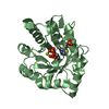
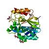
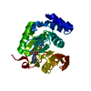
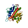
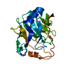
- PDB-2x5n: Crystal Structure of the SpRpn10 VWA domain -
+
Open data
ID or keywords:
Loading...
-
Basic information
Entry
Database: PDB / ID: 2x5n
Title
Crystal Structure of the SpRpn10 VWA domain
Components
26S PROTEASOME REGULATORY SUBUNIT RPN10
Keywords
NUCLEAR PROTEIN / NUCLEUS / UBIQUITIN / PROTEASOME
Function / homology
Function and homology information
stalled replication fork localization to nuclear periphery / mitotic recombination-dependent replication fork processing / proteasome regulatory particle, lid subcomplex / proteasome regulatory particle, base subcomplex / K48-linked polyubiquitin modification-dependent protein binding / proteasomal protein catabolic process / proteasome-mediated ubiquitin-dependent protein catabolic process / nucleus / cytosol Similarity search - Function
von Willebrand factor, type A domain / Proteasome subunit Rpn10 / von Willebrand factor type A domain / Ubiquitin interacting motif / Ubiquitin-interacting motif (UIM) domain profile. / VWFA domain profile. / von Willebrand factor (vWF) type A domain / von Willebrand factor, type A / von Willebrand factor A-like domain superfamily / Rossmann fold ...von Willebrand factor, type A domain / Proteasome subunit Rpn10 / von Willebrand factor type A domain / Ubiquitin interacting motif / Ubiquitin-interacting motif (UIM) domain profile. / VWFA domain profile. / von Willebrand factor (vWF) type A domain / von Willebrand factor, type A / von Willebrand factor A-like domain superfamily / Rossmann fold / 3-Layer(aba) Sandwich / Alpha Beta Similarity search - Domain/homology
Monochromator: DIAMOND (111), GE(220) / Protocol: SINGLE WAVELENGTH / Monochromatic (M) / Laue (L): M / Scattering type: x-ray
Radiation wavelength
Wavelength: 0.933 Å / Relative weight: 1
Reflection
Resolution: 1.27→56.1 Å / Num. obs: 38709 / % possible obs: 99.4 % / Observed criterion σ(I): 2 / Redundancy: 7 % / Biso Wilson estimate: 8.23 Å2 / Rmerge(I) obs: 0.04 / Net I/σ(I): 19.37
Reflection shell
Resolution: 1.27→1.4 Å / Redundancy: 6.8 % / Rmerge(I) obs: 0.16 / Mean I/σ(I) obs: 7.34 / % possible all: 98.7
-
Processing
Software
Name
Classification
MOSFLM
datareduction
SCALA
datascaling
SHELXD
phasing
SHELXE
phasing
PHENIX
refinement
Refinement
Method to determine structure: SAD Starting model: NONE Resolution: 1.3→27.738 Å / SU ML: 0.11 / Phase error: 13.88 / Stereochemistry target values: ML
Rfactor
Num. reflection
% reflection
Rfree
0.1646
1887
5 %
Rwork
0.1228
-
-
obs
0.1248
37864
97.83 %
Solvent computation
Shrinkage radii: 0.9 Å / VDW probe radii: 1.11 Å / Solvent model: FLAT BULK SOLVENT MODEL / Bsol: 41.953 Å2 / ksol: 0.458 e/Å3
Displacement parameters
Biso mean: 15.73 Å2
Baniso -1
Baniso -2
Baniso -3
1-
0.7849 Å2
-0 Å2
0.4916 Å2
2-
-
0.1402 Å2
0 Å2
3-
-
-
-0.9251 Å2
Refinement step
Cycle: LAST / Resolution: 1.3→27.738 Å
Protein
Nucleic acid
Ligand
Solvent
Total
Num. atoms
1469
0
15
271
1755
Refine LS restraints
Refine-ID
Type
Dev ideal
Number
X-RAY DIFFRACTION
f_bond_d
0.01
1637
X-RAY DIFFRACTION
f_angle_d
1.587
2237
X-RAY DIFFRACTION
f_dihedral_angle_d
16.794
627
X-RAY DIFFRACTION
f_chiral_restr
0.215
254
X-RAY DIFFRACTION
f_plane_restr
0.007
299
LS refinement shell
Resolution (Å)
Rfactor Rfree
Num. reflection Rfree
Rfactor Rwork
Num. reflection Rwork
Refine-ID
% reflection obs (%)
1.3-1.3465
0.1756
197
0.1199
3445
X-RAY DIFFRACTION
95
1.3465-1.4004
0.1899
185
0.1232
3542
X-RAY DIFFRACTION
96
1.4004-1.4641
0.173
165
0.1139
3546
X-RAY DIFFRACTION
97
1.4641-1.5413
0.1708
202
0.104
3567
X-RAY DIFFRACTION
98
1.5413-1.6379
0.1514
194
0.1034
3570
X-RAY DIFFRACTION
98
1.6379-1.7643
0.1518
205
0.1037
3621
X-RAY DIFFRACTION
99
1.7643-1.9418
0.1783
195
0.1109
3546
X-RAY DIFFRACTION
97
1.9418-2.2227
0.1444
169
0.1055
3663
X-RAY DIFFRACTION
99
2.2227-2.7999
0.1521
170
0.1179
3700
X-RAY DIFFRACTION
99
2.7999-27.7447
0.1651
205
0.1417
3777
X-RAY DIFFRACTION
100
+
About Yorodumi
-
News
-
Feb 9, 2022. New format data for meta-information of EMDB entries
New format data for meta-information of EMDB entries
Version 3 of the EMDB header file is now the official format.
The previous official version 1.9 will be removed from the archive.
In the structure databanks used in Yorodumi, some data are registered as the other names, "COVID-19 virus" and "2019-nCoV". Here are the details of the virus and the list of structure data.
Jan 31, 2019. EMDB accession codes are about to change! (news from PDBe EMDB page)
EMDB accession codes are about to change! (news from PDBe EMDB page)
The allocation of 4 digits for EMDB accession codes will soon come to an end. Whilst these codes will remain in use, new EMDB accession codes will include an additional digit and will expand incrementally as the available range of codes is exhausted. The current 4-digit format prefixed with “EMD-” (i.e. EMD-XXXX) will advance to a 5-digit format (i.e. EMD-XXXXX), and so on. It is currently estimated that the 4-digit codes will be depleted around Spring 2019, at which point the 5-digit format will come into force.
The EM Navigator/Yorodumi systems omit the EMD- prefix.
Related info.:Q: What is EMD? / ID/Accession-code notation in Yorodumi/EM Navigator
Yorodumi is a browser for structure data from EMDB, PDB, SASBDB, etc.
This page is also the successor to EM Navigator detail page, and also detail information page/front-end page for Omokage search.
The word "yorodu" (or yorozu) is an old Japanese word meaning "ten thousand". "mi" (miru) is to see.
Related info.:EMDB / PDB / SASBDB / Comparison of 3 databanks / Yorodumi Search / Aug 31, 2016. New EM Navigator & Yorodumi / Yorodumi Papers / Jmol/JSmol / Function and homology information / Changes in new EM Navigator and Yorodumi
 Movie
Movie Controller


 Open data
 Basic information
 Components
 Keywords
 Function and homology information
 X-RAY DIFFRACTION /
  Authors
 Citation
 Structure visualization
 Downloads & links
 Other downloads
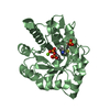

 PDBj
PDBj


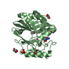
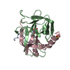
 Assembly
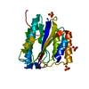


 Mass: 96.063 Da / Num. of mol.: 3 / Source method: obtained synthetically / Formula: SO4
 Mass: 18.015 Da / Num. of mol.: 271 / Source method: isolated from a natural source / Formula: H2O
 Sample preparation
 / Beamline: ID14-2 / Wavelength: 0.933
  Processing