 Movie
Movie Controller

[English] 日本語
 Yorodumi
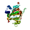
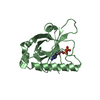
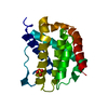
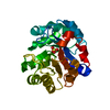
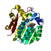
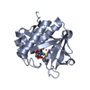
- PDB-1jm1: Crystal structure of the soluble domain of the Rieske protein II ... -
+ Open data

- Basic information
| Entry | Database: PDB / ID: 1jm1 | ||||||
|---|---|---|---|---|---|---|---|
| Title | Crystal structure of the soluble domain of the Rieske protein II (soxF) from Sulfolobus acidocaldarius | ||||||
 Components Components | Rieske iron-sulfur protein soxF | ||||||
 Keywords Keywords | electron transport / oxidoreductase / Rieske iron-sulfur protein / respiratory chain | ||||||
| Function / homology |  Function and homology information Function and homology information | ||||||
| Biological species |   Sulfolobus acidocaldarius (acidophilic) Sulfolobus acidocaldarius (acidophilic) | ||||||
| Method |  X-RAY DIFFRACTION / SYNCHROTRON / MAD / Resolution: 1.11 Å X-RAY DIFFRACTION / SYNCHROTRON / MAD / Resolution: 1.11 Å | ||||||
 Authors Authors | Boenisch, H. / Schmidt, C.L. / Schaefer, G. / Ladenstein, R. | ||||||
 Citation Citation | Journal: J.Mol.Biol. / Year: 2002 Title: The structure of the soluble domain of an archaeal Rieske iron-sulfur protein at 1.1 A resolution. Authors: Bonisch, H. / Schmidt, C.L. / Schafer, G. / Ladenstein, R. #1: Journal: Acta Crystallogr.,Sect.D / Year: 2000Title: Crystallization and preliminary crystallographic analysis of Rieske iron-sulfur protein II (soxF) from Sulfolobus acidocaldarius Authors: Boenisch, H. / Schmidt, C.L. / Schaefer, G. / Ladenstein, R. #2: Journal: Biochem.Biophys.Res.Commun. / Year: 1997Title: Expression of the Sulfolobus acidocaldarius Rieske iron sulfur protein II (soxF) with the correctly inserted [2Fe-2S] cluster in Escherichia coli Authors: Schmidt, C.L. / Hatzfeld, O.M. / Petersen, A. / Link, T.A. / Schaefer, G. | ||||||
| History |
|
- Structure visualization
| Structure viewer | Molecule: MolmilJmol/JSmol |
|---|
- Downloads & links
-Download
| PDBx/mmCIF format | 1jm1.cif.gz | 138.9 KB | Display | PDBx/mmCIF format |
|---|---|---|---|---|
| PDB format | pdb1jm1.ent.gz | 110.6 KB | Display | PDB format |
| PDBx/mmJSON format | 1jm1.json.gz | Tree view | PDBx/mmJSON format | |
| Others |  Other downloads Other downloads |
-Validation report
| Arichive directory | https://data.pdbj.org/pub/pdb/validation_reports/jm/1jm1ftp://data.pdbj.org/pub/pdb/validation_reports/jm/1jm1 | HTTPS FTP |
|---|
-Related structure data
| Similar structure data |
|---|

-Links
 PDBj
PDBj


- Assembly
| Deposited unit | 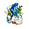
| ||||||||||
|---|---|---|---|---|---|---|---|---|---|---|---|
| 1 |
| ||||||||||
| Unit cell |
|
-Components
| #1: Protein | Mass: 21699.285 Da / Num. of mol.: 1 / Fragment: Soluble domain, C-terminal residues 47 - 250 Source method: isolated from a genetically manipulated source Source: (gene. exp.) Sulfolobus acidocaldarius (acidophilic)Gene: SOXF / Plasmid: pET3A / Species (production host): Escherichia coli / Production host:  |
|---|---|
| #2: Chemical | ChemComp-MG /   Mass: 24.305 Da / Num. of mol.: 1 / Source method: obtained synthetically / Formula: Mg Mass: 24.305 Da / Num. of mol.: 1 / Source method: obtained synthetically / Formula: Mg |
| #3: Chemical | ChemComp-FES /   Mass: 175.820 Da / Num. of mol.: 1 / Source method: obtained synthetically / Formula: Fe2S2 Mass: 175.820 Da / Num. of mol.: 1 / Source method: obtained synthetically / Formula: Fe2S2 |
| #4: Water | ChemComp-HOH /  Mass: 18.015 Da / Num. of mol.: 285 / Source method: isolated from a natural source / Formula: H2O Mass: 18.015 Da / Num. of mol.: 285 / Source method: isolated from a natural source / Formula: H2O |
| Has protein modification | Y |
-Experimental details
-Experiment
| Experiment | Method: X-RAY DIFFRACTION / Number of used crystals: 1 |
|---|
- Sample preparation
| Crystal | Density Matthews: 3.24 Å3/Da / Density % sol: 62 % | ||||||||||||||||||||||||
|---|---|---|---|---|---|---|---|---|---|---|---|---|---|---|---|---|---|---|---|---|---|---|---|---|---|
| Crystal grow | Temperature: 293 K / Method: vapor diffusion, sitting drop / pH: 7 Details: PEG 8000, magnesium acetate, sodium cacodylate, pH 7.0, VAPOR DIFFUSION, SITTING DROP, at 293K | ||||||||||||||||||||||||
| Crystal grow | *PLUS | ||||||||||||||||||||||||
| Components of the solutions | *PLUS
|
-Data collection
| Diffraction | Mean temperature: 100 K |
|---|---|
| Diffraction source | Source: SYNCHROTRON / Site: EMBL/DESY, HAMBURG  / Beamline: BW7A / Wavelength: 1 Å / Beamline: BW7A / Wavelength: 1 Å |
| Detector | Type: MARRESEARCH / Detector: CCD / Date: Oct 29, 1999 |
| Radiation | Protocol: MAD / Monochromatic (M) / Laue (L): M / Scattering type: x-ray |
| Radiation wavelength | Wavelength: 1 Å / Relative weight: 1 |
| Reflection | Resolution: 1.11→20 Å / Num. all: 112015 / Num. obs: 109985 / % possible obs: 98.2 % / Observed criterion σ(F): 0 / Observed criterion σ(I): -3 / Redundancy: 18.2 % / Biso Wilson estimate: 12.2 Å2 / Rmerge(I) obs: 0.046 / Net I/σ(I): 32.4 |
| Reflection shell | Resolution: 1.11→1.13 Å / Rmerge(I) obs: 0.275 / Mean I/σ(I) obs: 3.8 / Num. unique all: 4935 / % possible all: 73.6 |
| Reflection | *PLUS Lowest resolution: 69 Å / Num. obs: 112015 / Num. measured all: 2039387 / Rmerge(I) obs: 0.047 |
| Reflection shell | *PLUS % possible obs: 73.6 % |
- Processing
| Software |
| |||||||||||||||||||||||||
|---|---|---|---|---|---|---|---|---|---|---|---|---|---|---|---|---|---|---|---|---|---|---|---|---|---|---|
| Refinement | Method to determine structure: MAD / Resolution: 1.11→20 Å / Cross valid method: FREE R / σ(F): 0 / Stereochemistry target values: ENGH & HUBER
| |||||||||||||||||||||||||
| Displacement parameters | Biso mean: 19.64 Å2 | |||||||||||||||||||||||||
| Refinement step | Cycle: LAST / Resolution: 1.11→20 Å
| |||||||||||||||||||||||||
| Refine LS restraints |
| |||||||||||||||||||||||||
| Software | *PLUS Name: SHELXL / Version: 97 / Classification: refinement | |||||||||||||||||||||||||
| Refinement | *PLUS Lowest resolution: 69 Å | |||||||||||||||||||||||||
| Solvent computation | *PLUS | |||||||||||||||||||||||||
| Displacement parameters | *PLUS |
