解像度: 1.72→19.69 Å / Cor.coef. Fo:Fc: 0.969 / Cor.coef. Fo:Fc free: 0.955 / SU B: 2.465 / SU ML: 0.08 / TLS residual ADP flag: LIKELY RESIDUAL / 交差検証法: THROUGHOUT / σ(F): -3 / ESU R: 0.119 / ESU R Free: 0.116 / 立体化学のターゲット値: MAXIMUM LIKELIHOOD 詳細: HYDROGENS HAVE BEEN ADDED IN THE RIDING POSITIONS. NO REFLECTIONS WERE EXCLUDED ON THE BASIS OF A SIGMA CUTOFF
Rfactor
反射数
%反射
Selection details
Rfree
0.20809
965
5.2 %
RANDOM
Rwork
0.16579
-
-
-
all
0.16791
17769
-
-
obs
0.16791
17769
97.34 %
-
溶媒の処理
イオンプローブ半径: 0.8 Å / 減衰半径: 0.8 Å / VDWプローブ半径: 1.4 Å / 溶媒モデル: BABINET MODEL WITH MASK
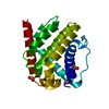
 ムービー
ムービー コントローラー


 データを開く
 基本情報
基本情報 要素
要素 キーワード
キーワード 機能・相同性情報
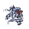
 X線回折 /
  データ登録者
 引用
引用 構造の表示
構造の表示 ダウンロードとリンク
 その他のダウンロード
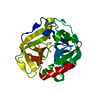

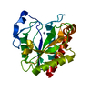


 PDBj
PDBj 集合体
集合体


 分子量: 118.174 Da / 分子数: 1 / 由来タイプ: 合成 / 式: C6H14O2 / コメント: 沈殿剤*YM
 分子量: 18.015 Da / 分子数: 124 / 由来タイプ: 天然 / 式: H2O
 試料調製
試料調製 / ビームライン: X11 / 波長: 1.1 Å
 解析
解析