 Movie
Movie Controller

[English] 日本語
 Yorodumi
- PDB-2y0o: The structure of a D-lyxose isomerase from the sigmaB regulon of ... -
+ Open data

- Basic information
| Entry | Database: PDB / ID: 2y0o | ||||||
|---|---|---|---|---|---|---|---|
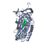
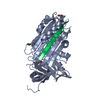
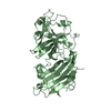
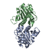
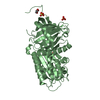
| Title | The structure of a D-lyxose isomerase from the sigmaB regulon of Bacillus subtilis | ||||||
 Components Components | PROBABLE D-LYXOSE KETOL-ISOMERASE | ||||||
 Keywords Keywords | ISOMERASE / CARBOHYDRATE METABOLISM / METAL-BINDING / SUGAR ISOMERASE / STRESS RESPONSE | ||||||
| Function / homology |  Function and homology information Function and homology informationD-lyxose ketol-isomerase / D-lyxose ketol-isomerase activity / metal ion binding Similarity search - Function | ||||||
| Biological species |  | ||||||
| Method |  X-RAY DIFFRACTION / SYNCHROTRON / MAD / Resolution: 1.229 Å X-RAY DIFFRACTION / SYNCHROTRON / MAD / Resolution: 1.229 Å | ||||||
 Authors Authors | Marles-Wright, J. / Lewis, R.J. | ||||||
 Citation Citation | Journal: Proteins / Year: 2011 Title: The Structure of a D-Lyxose Isomerase from the Sigmab Regulon of Bacillus Subtilis Authors: Marles-Wright, J. / Lewis, R.J. | ||||||
| History |
|
- Structure visualization
| Structure viewer | Molecule: MolmilJmol/JSmol |
|---|
- Downloads & links
-Download
| PDBx/mmCIF format | 2y0o.cif.gz | 96.9 KB | Display | PDBx/mmCIF format |
|---|---|---|---|---|
| PDB format | pdb2y0o.ent.gz | 73.8 KB | Display | PDB format |
| PDBx/mmJSON format | 2y0o.json.gz | Tree view | PDBx/mmJSON format | |
| Others |  Other downloads Other downloads |
-Validation report
| Arichive directory | https://data.pdbj.org/pub/pdb/validation_reports/y0/2y0oftp://data.pdbj.org/pub/pdb/validation_reports/y0/2y0o | HTTPS FTP |
|---|
-Related structure data
| Similar structure data |
|---|


-Links
 PDBj
PDBj- Assembly
| Deposited unit | 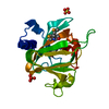
| |||||||||
|---|---|---|---|---|---|---|---|---|---|---|
| 1 | 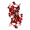
| |||||||||
| Unit cell |
| |||||||||
| Components on special symmetry positions |
|
-Components
| #1: Protein | Mass: 20487.422 Da / Num. of mol.: 1 Source method: isolated from a genetically manipulated source Details: ZINC AND ARSENIC COORDINATION IN ACTIVE SITE Source: (gene. exp.) Strain: 168 / Plasmid: PET28B / Production host: | ||||||
|---|---|---|---|---|---|---|---|
| #2: Chemical | ChemComp-ZN /   Mass: 65.409 Da / Num. of mol.: 1 / Source method: obtained synthetically / Formula: Zn Mass: 65.409 Da / Num. of mol.: 1 / Source method: obtained synthetically / Formula: Zn | ||||||
| #3: Chemical |   Mass: 96.063 Da / Num. of mol.: 2 / Source method: obtained synthetically / Formula: SO4 Mass: 96.063 Da / Num. of mol.: 2 / Source method: obtained synthetically / Formula: SO4#4: Chemical | ChemComp-ARS / |   Mass: 74.922 Da / Num. of mol.: 1 / Source method: obtained synthetically / Formula: As Mass: 74.922 Da / Num. of mol.: 1 / Source method: obtained synthetically / Formula: As#5: Water | ChemComp-HOH / |  Mass: 18.015 Da / Num. of mol.: 268 / Source method: isolated from a natural source / Formula: H2O Mass: 18.015 Da / Num. of mol.: 268 / Source method: isolated from a natural source / Formula: H2OHas protein modification | Y | |
-Experimental details
-Experiment
| Experiment | Method: X-RAY DIFFRACTION / Number of used crystals: 1 |
|---|
- Sample preparation
| Crystal | Density Matthews: 2.21 Å3/Da / Density % sol: 44.4 % / Description: NONE |
|---|---|
| Crystal grow | pH: 6.5 Details: 0.1 M SODIUM CACODYLATE PH 6.5 0.2 M NACL 2.0 M AMMONIUM SULFATE 1UL:1UL DROPS WITH PROTEIN IN WATER AT 11.5 MG/ML |
-Data collection
| Diffraction | Mean temperature: 93 K |
|---|---|
| Diffraction source | Source: SYNCHROTRON / Site: ESRF  / Beamline: ID29 / Wavelength: 0.9793 / Beamline: ID29 / Wavelength: 0.9793 |
| Detector | Type: ADSC CCD / Detector: CCD / Date: Apr 24, 2010 / Details: MIRRORS |
| Radiation | Protocol: MAD / Monochromatic (M) / Laue (L): M / Scattering type: x-ray |
| Radiation wavelength | Wavelength: 0.9793 Å / Relative weight: 1 |
| Reflection | Resolution: 1.23→35.45 Å / Num. obs: 51297 / % possible obs: 99.4 % / Observed criterion σ(I): 2 / Redundancy: 8.9 % / Biso Wilson estimate: 12.9 Å2 / Rmerge(I) obs: 0.08 / Net I/σ(I): 13.8 |
| Reflection shell | Resolution: 1.23→1.3 Å / Redundancy: 2.6 % / Rmerge(I) obs: 0.37 / Mean I/σ(I) obs: 2.6 / % possible all: 98.9 |
- Processing
| Software |
| |||||||||||||||||||||||||||||||||||||||||||||||||||||||||||||||||||||||||||||
|---|---|---|---|---|---|---|---|---|---|---|---|---|---|---|---|---|---|---|---|---|---|---|---|---|---|---|---|---|---|---|---|---|---|---|---|---|---|---|---|---|---|---|---|---|---|---|---|---|---|---|---|---|---|---|---|---|---|---|---|---|---|---|---|---|---|---|---|---|---|---|---|---|---|---|---|---|---|---|
| Refinement | Method to determine structure: MAD Starting model: NONE Resolution: 1.229→34.872 Å / SU ML: 0.17 / σ(F): 1.37 / Phase error: 16.14 / Stereochemistry target values: ML
| |||||||||||||||||||||||||||||||||||||||||||||||||||||||||||||||||||||||||||||
| Solvent computation | Shrinkage radii: 0.83 Å / VDW probe radii: 1.1 Å / Solvent model: FLAT BULK SOLVENT MODEL / Bsol: 40.657 Å2 / ksol: 0.375 e/Å3 | |||||||||||||||||||||||||||||||||||||||||||||||||||||||||||||||||||||||||||||
| Displacement parameters | Biso mean: 18.91 Å2
| |||||||||||||||||||||||||||||||||||||||||||||||||||||||||||||||||||||||||||||
| Refinement step | Cycle: LAST / Resolution: 1.229→34.872 Å
| |||||||||||||||||||||||||||||||||||||||||||||||||||||||||||||||||||||||||||||
| Refine LS restraints |
| |||||||||||||||||||||||||||||||||||||||||||||||||||||||||||||||||||||||||||||
| LS refinement shell |
|
