 Movie
Movie Controller

[English] 日本語
 Yorodumi
- PDB-4dsr: Crystal structure of peroxiredoxin Ahp1 from Saccharomyces cerevi... -
+ Open data

- Basic information
| Entry | Database: PDB / ID: 4dsr | ||||||
|---|---|---|---|---|---|---|---|
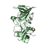
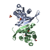
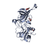
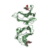
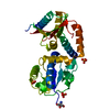
| Title | Crystal structure of peroxiredoxin Ahp1 from Saccharomyces cerevisiae in reduced form | ||||||
 Components Components | Peroxiredoxin type-2 | ||||||
 Keywords Keywords | OXIDOREDUCTASE / PEROXIREDOXIN / Peroxidase / Thioredoxin-like Fold / Alkyl hydroperoxide reductase | ||||||
| Function / homology |  Function and homology information Function and homology informationTP53 Regulates Metabolic Genes / Detoxification of Reactive Oxygen Species / thioredoxin-dependent peroxiredoxin / thioredoxin peroxidase activity / response to metal ion / cell redox homeostasis / hydrogen peroxide catabolic process / peroxisome / cellular response to oxidative stress / mitochondrion ...TP53 Regulates Metabolic Genes / Detoxification of Reactive Oxygen Species / thioredoxin-dependent peroxiredoxin / thioredoxin peroxidase activity / response to metal ion / cell redox homeostasis / hydrogen peroxide catabolic process / peroxisome / cellular response to oxidative stress / mitochondrion / plasma membrane / cytosol / cytoplasm Similarity search - Function | ||||||
| Biological species |  | ||||||
| Method |  X-RAY DIFFRACTION / SYNCHROTRON / MOLECULAR REPLACEMENT / Resolution: 2.91 Å X-RAY DIFFRACTION / SYNCHROTRON / MOLECULAR REPLACEMENT / Resolution: 2.91 Å | ||||||
 Authors Authors | Lian, F.M. / Yu, J. / Ma, X.X. / Yu, X.J. / Chen, Y. / Zhou, C.Z. | ||||||
 Citation Citation | Journal: J.Biol.Chem. / Year: 2012 Title: Structural Snapshots of Yeast Alkyl Hydroperoxide Reductase Ahp1 Peroxiredoxin Reveal a Novel Two-cysteine Mechanism of Electron Transfer to Eliminate Reactive Oxygen Species. Authors: Lian, F.M. / Yu, J. / Ma, X.X. / Yu, X.J. / Chen, Y. / Zhou, C.Z. | ||||||
| History |
|
- Structure visualization
| Structure viewer | Molecule: MolmilJmol/JSmol |
|---|
- Downloads & links
-Download
| PDBx/mmCIF format | 4dsr.cif.gz | 138 KB | Display | PDBx/mmCIF format |
|---|---|---|---|---|
| PDB format | pdb4dsr.ent.gz | 109.3 KB | Display | PDB format |
| PDBx/mmJSON format | 4dsr.json.gz | Tree view | PDBx/mmJSON format | |
| Others |  Other downloads Other downloads |
-Validation report
| Arichive directory | https://data.pdbj.org/pub/pdb/validation_reports/ds/4dsrftp://data.pdbj.org/pub/pdb/validation_reports/ds/4dsr | HTTPS FTP |
|---|
-Related structure data
| Related structure data |  4dsqC  4dssC  1tp9S C: citing same article ( S: Starting model for refinement |
|---|---|
| Similar structure data |


-Links
 PDBj
PDBj
- Assembly
| Deposited unit | 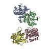
| ||||||||
|---|---|---|---|---|---|---|---|---|---|
| 1 | 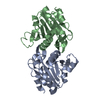
| ||||||||
| 2 | 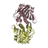
| ||||||||
| Unit cell |
|
-Components
| #1: Protein | Mass: 20149.730 Da / Num. of mol.: 4 Source method: isolated from a genetically manipulated source Source: (gene. exp.) Strain: S288C / Gene: AHP1, L2916, L9354.5, YLR109W / Plasmid: pET28a-derived / Production host:  #2: Water | ChemComp-HOH / |  Mass: 18.015 Da / Num. of mol.: 11 / Source method: isolated from a natural source / Formula: H2O Mass: 18.015 Da / Num. of mol.: 11 / Source method: isolated from a natural source / Formula: H2O |
|---|
-Experimental details
-Experiment
| Experiment | Method: X-RAY DIFFRACTION / Number of used crystals: 1 |
|---|
- Sample preparation
| Crystal | Density Matthews: 2.59 Å3/Da / Density % sol: 52.53 % |
|---|---|
| Crystal grow | Temperature: 289 K / Method: vapor diffusion, hanging drop / pH: 7.5 Details: 25% polyethylene glycol 3350, 0.2M ammonium sulfate, 0.1M HEPES-NaOH, pH 7.5, VAPOR DIFFUSION, HANGING DROP, temperature 289K |
-Data collection
| Diffraction | Mean temperature: 100 K |
|---|---|
| Diffraction source | Source: SYNCHROTRON / Site: SSRF  / Beamline: BL17U / Wavelength: 0.9791 Å / Beamline: BL17U / Wavelength: 0.9791 Å |
| Detector | Type: ADSC QUANTUM 315r / Detector: CCD / Date: Apr 20, 2011 |
| Radiation | Protocol: SINGLE WAVELENGTH / Monochromatic (M) / Laue (L): M / Scattering type: x-ray |
| Radiation wavelength | Wavelength: 0.9791 Å / Relative weight: 1 |
| Reflection | Resolution: 2.91→50 Å / Num. all: 16465 / Num. obs: 16465 / % possible obs: 91.3 % / Observed criterion σ(F): 0 / Observed criterion σ(I): -3 / Redundancy: 4 % / Biso Wilson estimate: 70.2 Å2 / Rmerge(I) obs: 0.131 / Rsym value: 0.131 / Net I/σ(I): 7.68 |
| Reflection shell | Resolution: 2.91→3.01 Å / Redundancy: 3.4 % / Rmerge(I) obs: 0.369 / Mean I/σ(I) obs: 2.991 / Num. unique all: 1642 / Rsym value: 0.369 / % possible all: 91.5 |
- Processing
| Software |
| |||||||||||||||||||||||||||||||||||||||||||||||||||||||||||||||||
|---|---|---|---|---|---|---|---|---|---|---|---|---|---|---|---|---|---|---|---|---|---|---|---|---|---|---|---|---|---|---|---|---|---|---|---|---|---|---|---|---|---|---|---|---|---|---|---|---|---|---|---|---|---|---|---|---|---|---|---|---|---|---|---|---|---|---|
| Refinement | Method to determine structure: MOLECULAR REPLACEMENT Starting model: PDB ENTRY 1TP9 Resolution: 2.91→44.2 Å / Cor.coef. Fo:Fc: 0.903 / Cor.coef. Fo:Fc free: 0.883 / Cross valid method: THROUGHOUT / ESU R Free: 0.494 / Stereochemistry target values: MAXIMUM LIKELIHOOD / Details: HYDROGENS HAVE BEEN ADDED IN THE RIDING POSITIONS
| |||||||||||||||||||||||||||||||||||||||||||||||||||||||||||||||||
| Solvent computation | Ion probe radii: 0.8 Å / Shrinkage radii: 0.8 Å / VDW probe radii: 1.4 Å / Solvent model: MASK | |||||||||||||||||||||||||||||||||||||||||||||||||||||||||||||||||
| Displacement parameters | Biso mean: 62.23 Å2
| |||||||||||||||||||||||||||||||||||||||||||||||||||||||||||||||||
| Refinement step | Cycle: LAST / Resolution: 2.91→44.2 Å
| |||||||||||||||||||||||||||||||||||||||||||||||||||||||||||||||||
| Refine LS restraints |
| |||||||||||||||||||||||||||||||||||||||||||||||||||||||||||||||||
| LS refinement shell | Resolution: 2.91→2.986 Å / Total num. of bins used: 20
|
