 Movie
Movie Controller


+ Open data

- Basic information
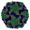
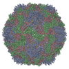
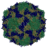
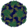
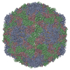
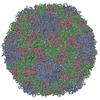
| Entry | Database: PDB / ID: 6rjf | |||||||||||||||
|---|---|---|---|---|---|---|---|---|---|---|---|---|---|---|---|---|
| Title | Echovirus 1 intact particle | |||||||||||||||
 Components Components |
| |||||||||||||||
 Keywords Keywords | VIRUS / VIRAL COAT PROTEIN / CAPSID / PICORNAVIRUS / ECHOVIRUS / ICOSAHEDRAL VIRUS | |||||||||||||||
| Function / homology |  Function and homology information Function and homology informationcaveolin-mediated endocytosis of virus by host cell / symbiont-mediated suppression of host cytoplasmic pattern recognition receptor signaling pathway via inhibition of RIG-I activity / picornain 2A / symbiont-mediated suppression of host mRNA export from nucleus / symbiont genome entry into host cell via pore formation in plasma membrane / picornain 3C / T=pseudo3 icosahedral viral capsid / host cell cytoplasmic vesicle membrane / viral capsid / ribonucleoside triphosphate phosphatase activity ...caveolin-mediated endocytosis of virus by host cell / symbiont-mediated suppression of host cytoplasmic pattern recognition receptor signaling pathway via inhibition of RIG-I activity / picornain 2A / symbiont-mediated suppression of host mRNA export from nucleus / symbiont genome entry into host cell via pore formation in plasma membrane / picornain 3C / T=pseudo3 icosahedral viral capsid / host cell cytoplasmic vesicle membrane / viral capsid / ribonucleoside triphosphate phosphatase activity / nucleoside-triphosphate phosphatase / channel activity / monoatomic ion transmembrane transport / host cell cytoplasm / DNA replication / RNA helicase activity / symbiont-mediated suppression of host gene expression / symbiont-mediated activation of host autophagy / RNA-directed RNA polymerase / cysteine-type endopeptidase activity / viral RNA genome replication / RNA-directed RNA polymerase activity / virion attachment to host cell / host cell nucleus / structural molecule activity / DNA-templated transcription / proteolysis / RNA binding / zinc ion binding / ATP binding / cytoplasm Similarity search - Function | |||||||||||||||
| Biological species |  Echovirus E1 Echovirus E1 | |||||||||||||||
| Method | ELECTRON MICROSCOPY / single particle reconstruction / cryo EM / Resolution: 3.5 Å | |||||||||||||||
 Authors Authors | Domanska, A. / Ruokolainen, V.P. / Pelliccia, M. / Laajala, M.A. / Marjomaki, V.S. / Butcher, S.J. | |||||||||||||||
| Funding support |  Finland, 4items Finland, 4items
| |||||||||||||||
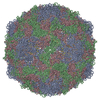
 Citation Citation | Journal: J Virol / Year: 2019 Title: Extracellular Albumin and Endosomal Ions Prime Enterovirus Particles for Uncoating That Can Be Prevented by Fatty Acid Saturation. Authors: Visa Ruokolainen / Aušra Domanska / Mira Laajala / Maria Pelliccia / Sarah J Butcher / Varpu Marjomäki /  Abstract: There is limited information about the molecular triggers leading to the uncoating of enteroviruses under physiological conditions. Using real-time spectroscopy and sucrose gradients with ...There is limited information about the molecular triggers leading to the uncoating of enteroviruses under physiological conditions. Using real-time spectroscopy and sucrose gradients with radioactively labeled virus, we show at 37°C, the formation of albumin-triggered, metastable uncoating intermediate of echovirus 1 without receptor engagement. This conversion was blocked by saturating the albumin with fatty acids. High potassium but low sodium and calcium concentrations, mimicking the endosomal environment, also induced the formation of a metastable uncoating intermediate of echovirus 1. Together, these factors boosted the formation of the uncoating intermediate, and the infectivity of this intermediate was retained, as judged by end-point titration. Cryo-electron microscopy reconstruction of the virions treated with albumin and high potassium, low sodium, and low calcium concentrations resulted in a 3.6-Å resolution model revealing a fenestrated capsid showing 4% expansion and loss of the pocket factor, similarly to altered (A) particles described for other enteroviruses. The dimer interface between VP2 molecules was opened, the VP1 N termini disordered and most likely externalized. The RNA was clearly visible, anchored to the capsid. The results presented here suggest that extracellular albumin, partially saturated with fatty acids, likely leads to the formation of the infectious uncoating intermediate prior to the engagement with the cellular receptor. In addition, changes in mono- and divalent cations, likely occurring in endosomes, promote capsid opening and genome release. There is limited information about the uncoating of enteroviruses under physiological conditions. Here, we focused on physiologically relevant factors that likely contribute to opening of echovirus 1 and other B-group enteroviruses. By combining biochemical and structural data, we show that, before entering cells, extracellular albumin is capable of priming the virus into a metastable yet infectious intermediate state. The ionic changes that are suggested to occur in endosomes can further contribute to uncoating and promote genome release, once the viral particle is endocytosed. Importantly, we provide a detailed high-resolution structure of a virion after treatment with albumin and a preset ion composition, showing pocket factor release, capsid expansion, and fenestration and the clearly visible genome still anchored to the capsid. This study provides valuable information about the physiological factors that contribute to the opening of B group enteroviruses. #1: Journal: Acta Crystallogr D Biol Crystallogr / Year: 1998Title: Structure determination of echovirus 1. Authors: D J Filman / M W Wien / J A Cunningham / J M Bergelson / J M Hogle /  Abstract: The atomic structure of echovirus 1 (a member of the enterovirus genus of the picornavirus family) has been determined using cryo-crystallography and refined to 3.55 A resolution. Echovirus 1 ...The atomic structure of echovirus 1 (a member of the enterovirus genus of the picornavirus family) has been determined using cryo-crystallography and refined to 3.55 A resolution. Echovirus 1 crystallizes in space group P22121 with a = 352.45, b = 472.15 and c = 483.20 A. The crystals contain one full virus particle in the asymmetric unit allowing for 60-fold noncrystallographic symmetry averaging. The diffraction pattern shows strong pseudo-B-centering with reflections with h + l = 2n + 1 being systematically weak or absent below about 6 A resolution. The size of the unit cell and presence of pseudo-B-centering placed strong constraints on the allowed packing of the icosahedral particle in the crystal lattice. These constraints greatly facilitated the determination of the orientation and position of the virus by reducing the dimensionality of the search, but interactions between the crystallographic and noncrystallographic symmetries rendered the choice of space group ambiguous until very late in the structure determination. This structure determination provides a striking example of the power of packing analysis in molecular replacement and illustrates how subtle interactions between crystallographic and noncrystallographic symmetries can be resolved. #2: Journal: Acta Crystallogr D Biol Crystallogr / Year: 1996 Title: A pseudo-cell based approach to efficient crystallographic refinement of viruses. Authors: D H Jacobson / J M Hogle / D J Filman / Abstract: Strategies have been developed for the inexpensive refinement of atomic models of viruses and of other highly symmetric structures. These methods, which have been used in the refinement of several ...Strategies have been developed for the inexpensive refinement of atomic models of viruses and of other highly symmetric structures. These methods, which have been used in the refinement of several strains of poliovirus, focus on an arbitrary-sized parallelepiped (termed the 'protomer' box) containing a single complete averaged copy of the structural motif which forms the protein capsid, together with the fragments of other symmetry-related copies of the motif which are located in its immediate neighborhood. The Fourier transform of the protomer box provides reference structure factors for stereochemically restrained crystallographic refinement of the atomic model parameters. The phases of the reference structure factors are based on the averaged map, and are not permitted to change during the refinement. It is demonstrated that models refined using the protomer box methods do not differ significantly from models refined by more expensive full-cell calculations. | |||||||||||||||
| History |
|
- Structure visualization
| Movie |
Movie viewer |
|---|---|
| Structure viewer | Molecule: MolmilJmol/JSmol |
- Downloads & links
-Download
| PDBx/mmCIF format | 6rjf.cif.gz | 275.1 KB | Display | PDBx/mmCIF format |
|---|---|---|---|---|
| PDB format | pdb6rjf.ent.gz | 219.9 KB | Display | PDB format |
| PDBx/mmJSON format | 6rjf.json.gz | Tree view | PDBx/mmJSON format | |
| Others |  Other downloads Other downloads |
-Validation report
| Arichive directory | https://data.pdbj.org/pub/pdb/validation_reports/rj/6rjfftp://data.pdbj.org/pub/pdb/validation_reports/rj/6rjf | HTTPS FTP |
|---|
-Related structure data
| Related structure data | 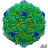 4903MC 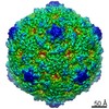 0565C  6o06C M: map data used to model this data C: citing same article ( |
|---|---|
| Similar structure data | |
| EM raw data | EMPIAR-10284 (Title: Extracellular albumin and endosomal ions prime enterovirus particles for uncoating that can be prevented by fatty acid saturation Data size: 2.4 TB Data #1: Unaligned multi-frame micrographs of control Echovirus 1 [micrographs - multiframe] Data #2: Unaligned multi-frame micrographs of control Echovirus 1 [micrographs - multiframe] Data #3: Unaligned multi-frame micrographs of treated Echovirus 1 (expanded particle) [micrographs - multiframe]) |


-Links
 PDBj
PDBj


- Assembly
| Deposited unit | 
|
|---|---|
| 1 | x 60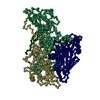
|
-Components
| #1: Protein | Mass: 31604.373 Da / Num. of mol.: 1 / Source method: isolated from a natural source / Source: (natural) Echovirus E1 / Strain: Human/Egypt/Farouk/1951 / References: UniProt: O91734 |
|---|---|
| #2: Protein | Mass: 28872.260 Da / Num. of mol.: 1 / Source method: isolated from a natural source / Source: (natural) Echovirus E1 / References: UniProt: O91734 |
| #3: Protein | Mass: 26471.074 Da / Num. of mol.: 1 / Source method: isolated from a natural source / Source: (natural) Echovirus E1 / References: UniProt: O91734 |
| #4: Protein | Mass: 7398.131 Da / Num. of mol.: 1 / Source method: isolated from a natural source / Source: (natural) Echovirus E1 / References: UniProt: C4B607*PLUS |
| #5: Chemical | ChemComp-PLM /   Mass: 256.424 Da / Num. of mol.: 1 / Source method: obtained synthetically / Formula: C16H32O2 Mass: 256.424 Da / Num. of mol.: 1 / Source method: obtained synthetically / Formula: C16H32O2 |
-Experimental details
-Experiment
| Experiment | Method: ELECTRON MICROSCOPY |
|---|---|
| EM experiment | Aggregation state: PARTICLE / 3D reconstruction method: single particle reconstruction |
- Sample preparation
| Component | Name: Echovirus E1 / Type: VIRUS / Entity ID: #1-#4 / Source: NATURAL |
|---|---|
| Molecular weight | Units: MEGADALTONS / Experimental value: NO |
| Source (natural) | Organism: Echovirus E1 |
| Details of virus | Empty: NO / Enveloped: NO / Isolate: OTHER / Type: VIRION |
| Natural host | Organism: Homo sapiens |
| Virus shell | Name: icosahedral / Diameter: 300 nm / Triangulation number (T number): 1 |
| Buffer solution | pH: 7.2 / Details: 2 mM magnesium chloride in PBS |
| Specimen | Embedding applied: NO / Shadowing applied: NO / Staining applied: NO / Vitrification applied: YES |
| Specimen support | Grid type: Quantifoil R2/2 |
| Vitrification | Instrument: HOMEMADE PLUNGER / Cryogen name: ETHANE |
- Electron microscopy imaging
| Experimental equipment |  Model: Talos Arctica / Image courtesy: FEI Company |
|---|---|
| Microscopy | Model: FEI TALOS ARCTICA |
| Electron gun | Electron source:  FIELD EMISSION GUN / Accelerating voltage: 200 kV / Illumination mode: FLOOD BEAM FIELD EMISSION GUN / Accelerating voltage: 200 kV / Illumination mode: FLOOD BEAM |
| Electron lens | Mode: BRIGHT FIELD |
| Specimen holder | Cryogen: NITROGEN |
| Image recording | Electron dose: 30 e/Å2 / Film or detector model: FEI FALCON III (4k x 4k) / Num. of real images: 979 |
- Processing
| EM software |
| ||||||||||||||||||||||||||||||||||||||||||||||||||||||||||||
|---|---|---|---|---|---|---|---|---|---|---|---|---|---|---|---|---|---|---|---|---|---|---|---|---|---|---|---|---|---|---|---|---|---|---|---|---|---|---|---|---|---|---|---|---|---|---|---|---|---|---|---|---|---|---|---|---|---|---|---|---|---|
| CTF correction | Type: PHASE FLIPPING AND AMPLITUDE CORRECTION | ||||||||||||||||||||||||||||||||||||||||||||||||||||||||||||
| Particle selection | Num. of particles selected: 45309 | ||||||||||||||||||||||||||||||||||||||||||||||||||||||||||||
| Symmetry | Point symmetry: I (icosahedral) | ||||||||||||||||||||||||||||||||||||||||||||||||||||||||||||
| 3D reconstruction | Resolution: 3.5 Å / Resolution method: FSC 0.143 CUT-OFF / Num. of particles: 45309 / Symmetry type: POINT | ||||||||||||||||||||||||||||||||||||||||||||||||||||||||||||
| Atomic model building |
| ||||||||||||||||||||||||||||||||||||||||||||||||||||||||||||
| Atomic model building |
|
