 Movie
Movie Controller


+ Open data

- Basic information
| Entry | Database: PDB / ID: 7dpf | ||||||
|---|---|---|---|---|---|---|---|
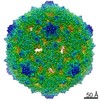
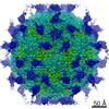
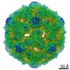
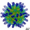
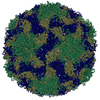
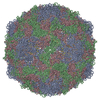
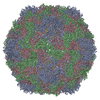
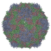
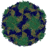
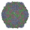
| Title | Cryo-EM structure of Coxsackievirus B1 mature virion | ||||||
 Components Components |
| ||||||
 Keywords Keywords | VIRUS / Coxsackievirus B1 / mature virion / Cryo-EM | ||||||
| Function / homology |  Function and homology information Function and homology informationsymbiont-mediated suppression of host cytoplasmic pattern recognition receptor signaling pathway via inhibition of RIG-I activity / picornain 2A / symbiont-mediated suppression of host mRNA export from nucleus / symbiont genome entry into host cell via pore formation in plasma membrane / picornain 3C / T=pseudo3 icosahedral viral capsid / host cell cytoplasmic vesicle membrane / viral capsid / ribonucleoside triphosphate phosphatase activity / host cell ...symbiont-mediated suppression of host cytoplasmic pattern recognition receptor signaling pathway via inhibition of RIG-I activity / picornain 2A / symbiont-mediated suppression of host mRNA export from nucleus / symbiont genome entry into host cell via pore formation in plasma membrane / picornain 3C / T=pseudo3 icosahedral viral capsid / host cell cytoplasmic vesicle membrane / viral capsid / ribonucleoside triphosphate phosphatase activity / host cell / nucleoside-triphosphate phosphatase / channel activity / monoatomic ion transmembrane transport / DNA replication / RNA helicase activity / endocytosis involved in viral entry into host cell / symbiont-mediated suppression of host gene expression / symbiont-mediated activation of host autophagy / RNA-directed RNA polymerase / cysteine-type endopeptidase activity / viral RNA genome replication / RNA-directed RNA polymerase activity / symbiont entry into host cell / virion attachment to host cell / host cell nucleus / DNA-templated transcription / structural molecule activity / proteolysis / RNA binding / zinc ion binding / ATP binding Similarity search - Function | ||||||
| Biological species |  Coxsackievirus B1 Coxsackievirus B1 | ||||||
| Method | ELECTRON MICROSCOPY / single particle reconstruction / cryo EM / Resolution: 3.2 Å | ||||||
 Authors Authors | Zheng, Q. / Li, S. | ||||||
 Citation Citation | Journal: Cell Host Microbe / Year: 2021 Title: Cryo-EM structures reveal the molecular basis of receptor-initiated coxsackievirus uncoating. Authors: Longfa Xu / Qingbing Zheng / Rui Zhu / Zhichao Yin / Hai Yu / Yu Lin / Yuanyuan Wu / Maozhou He / Yang Huang / Yichao Jiang / Hui Sun / Zhenghui Zha / Hongwei Yang / Qiongzi Huang / Dongqing ...Authors: Longfa Xu / Qingbing Zheng / Rui Zhu / Zhichao Yin / Hai Yu / Yu Lin / Yuanyuan Wu / Maozhou He / Yang Huang / Yichao Jiang / Hui Sun / Zhenghui Zha / Hongwei Yang / Qiongzi Huang / Dongqing Zhang / Zhenqin Chen / Xiangzhong Ye / Jinle Han / Lisheng Yang / Che Liu / Yuqiong Que / Mujin Fang / Ying Gu / Jun Zhang / Wenxin Luo / Z Hong Zhou / Shaowei Li / Tong Cheng / Ningshao Xia /   Abstract: Enterovirus uncoating receptors bind at the surface depression ("canyon") that encircles each capsid vertex causing the release of a host-derived lipid called "pocket factor" that is buried in a ...Enterovirus uncoating receptors bind at the surface depression ("canyon") that encircles each capsid vertex causing the release of a host-derived lipid called "pocket factor" that is buried in a hydrophobic pocket formed by the major viral capsid protein, VP1. Coxsackievirus and adenovirus receptor (CAR) is a universal uncoating receptor of group B coxsackieviruses (CVB). Here, we present five high-resolution cryoEM structures of CVB representing different stages of virus infection. Structural comparisons show that the CAR penetrates deeper into the canyon than other uncoating receptors, leading to a cascade of events: collapse of the VP1 hydrophobic pocket, high-efficiency release of the pocket factor and viral uncoating and genome release under neutral pH, as compared with low pH. Furthermore, we identified a potent therapeutic antibody that can neutralize viral infection by interfering with virion-CAR interactions, destabilizing the capsid and inducing virion disruption. Together, these results define the structural basis of CVB cell entry and antibody neutralization. | ||||||
| History |
|
- Structure visualization
| Movie |
Movie viewer |
|---|---|
| Structure viewer | Molecule: MolmilJmol/JSmol |
- Downloads & links
-Download
| PDBx/mmCIF format | 7dpf.cif.gz | 152.7 KB | Display | PDBx/mmCIF format |
|---|---|---|---|---|
| PDB format | pdb7dpf.ent.gz | 119.7 KB | Display | PDB format |
| PDBx/mmJSON format | 7dpf.json.gz | Tree view | PDBx/mmJSON format | |
| Others |  Other downloads Other downloads |
-Validation report
| Arichive directory | https://data.pdbj.org/pub/pdb/validation_reports/dp/7dpfftp://data.pdbj.org/pub/pdb/validation_reports/dp/7dpf | HTTPS FTP |
|---|
-Related structure data
| Related structure data | 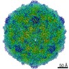 30805MC  7dpgC  7dpzC  7dq1C 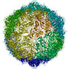 7dq4C 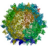 7dq7C M: map data used to model this data C: citing same article ( |
|---|---|
| Similar structure data |


-Links
 PDBj
PDBj


- Assembly
| Deposited unit | 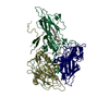
|
|---|---|
| 1 | x 60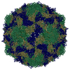
|
| 2 |
|
| 3 | x 5
|
| 4 | x 6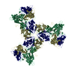
|
| 5 | 
|
| Symmetry | Point symmetry: (Schoenflies symbol: I (icosahedral)) |
-Components
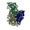
| #1: Protein | Mass: 31207.117 Da / Num. of mol.: 1 / Source method: isolated from a natural source / Source: (natural) Coxsackievirus B1 / References: UniProt: W8GTF7 |
|---|---|
| #2: Protein | Mass: 29122.744 Da / Num. of mol.: 1 / Source method: isolated from a natural source / Source: (natural) Coxsackievirus B1 / References: UniProt: A0A2S0RQC2 |
| #3: Protein | Mass: 26328.764 Da / Num. of mol.: 1 / Source method: isolated from a natural source / Source: (natural) Coxsackievirus B1 / References: UniProt: A0A0G4PYT0 |
| #4: Protein | Mass: 7484.246 Da / Num. of mol.: 1 / Source method: isolated from a natural source / Source: (natural) Coxsackievirus B1 / References: UniProt: A0A2S1FMR1 |
| #5: Chemical | ChemComp-PLM /   Mass: 256.424 Da / Num. of mol.: 1 / Source method: obtained synthetically / Formula: C16H32O2 / Feature type: SUBJECT OF INVESTIGATION Mass: 256.424 Da / Num. of mol.: 1 / Source method: obtained synthetically / Formula: C16H32O2 / Feature type: SUBJECT OF INVESTIGATION |
| Has ligand of interest | Y |
-Experimental details
-Experiment
| Experiment | Method: ELECTRON MICROSCOPY |
|---|---|
| EM experiment | Aggregation state: PARTICLE / 3D reconstruction method: single particle reconstruction |
- Sample preparation
| Component | Name: Coxsackievirus B1 / Type: VIRUS / Entity ID: #1-#4 / Source: NATURAL |
|---|---|
| Source (natural) | Organism: Coxsackievirus B1 |
| Details of virus | Empty: NO / Enveloped: NO / Isolate: STRAIN / Type: VIRION |
| Buffer solution | pH: 7.4 |
| Specimen | Embedding applied: NO / Shadowing applied: NO / Staining applied: NO / Vitrification applied: YES |
| Vitrification | Cryogen name: ETHANE |
- Electron microscopy imaging
| Experimental equipment |  Model: Tecnai F30 / Image courtesy: FEI Company |
|---|---|
| Microscopy | Model: FEI TECNAI F30 |
| Electron gun | Electron source:  FIELD EMISSION GUN / Accelerating voltage: 300 kV / Illumination mode: FLOOD BEAM FIELD EMISSION GUN / Accelerating voltage: 300 kV / Illumination mode: FLOOD BEAM |
| Electron lens | Mode: BRIGHT FIELD |
| Image recording | Electron dose: 25 e/Å2 / Detector mode: INTEGRATING / Film or detector model: FEI FALCON III (4k x 4k) |
- Processing
| CTF correction | Type: PHASE FLIPPING ONLY |
|---|---|
| 3D reconstruction | Resolution: 3.2 Å / Resolution method: FSC 0.143 CUT-OFF / Num. of particles: 35999 / Symmetry type: POINT |
