 Movie
Movie Controller

[English] 日本語
 Yorodumi
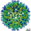
- EMDB-30813: Cryo-EM structure of Coxsackievirus B1 virion in complex with CAR... -
+ Open data

- Basic information
| Entry | Database: EMDB / ID: EMD-30813 | |||||||||
|---|---|---|---|---|---|---|---|---|---|---|
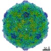
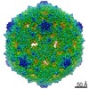
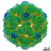
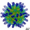
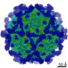
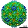
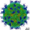
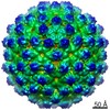
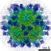
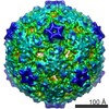
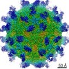
| Title | Cryo-EM structure of Coxsackievirus B1 virion in complex with CAR at physiological temperature | |||||||||
 Map data Map data | Cryo-EM structure of CAR triggered Coxsachievirus B1 A-particle | |||||||||
 Sample Sample |
| |||||||||
 Keywords Keywords | Coxsackievirus B1 / CAR / Cryo-EM / A-particle / VIRUS | |||||||||
| Function / homology |  Function and homology information Function and homology informationAV node cell-bundle of His cell adhesion involved in cell communication / cell adhesive protein binding involved in AV node cell-bundle of His cell communication / homotypic cell-cell adhesion / AV node cell to bundle of His cell communication / epithelial structure maintenance / regulation of AV node cell action potential / gamma-delta T cell activation / apicolateral plasma membrane / primordial germ cell migration / connexin binding ...AV node cell-bundle of His cell adhesion involved in cell communication / cell adhesive protein binding involved in AV node cell-bundle of His cell communication / homotypic cell-cell adhesion / AV node cell to bundle of His cell communication / epithelial structure maintenance / regulation of AV node cell action potential / gamma-delta T cell activation / apicolateral plasma membrane / primordial germ cell migration / connexin binding / cell-cell junction organization / transepithelial transport / heterophilic cell-cell adhesion / cardiac muscle cell development / symbiont-mediated suppression of host cytoplasmic pattern recognition receptor signaling pathway via inhibition of RIG-I activity / intercalated disc / bicellular tight junction / neutrophil chemotaxis / cell adhesion molecule binding / acrosomal vesicle / Cell surface interactions at the vascular wall / picornain 2A / filopodium / symbiont-mediated suppression of host mRNA export from nucleus / adherens junction / PDZ domain binding / neuromuscular junction / mitochondrion organization / symbiont genome entry into host cell via pore formation in plasma membrane / picornain 3C / T=pseudo3 icosahedral viral capsid / host cell cytoplasmic vesicle membrane / beta-catenin binding / integrin binding / Immunoregulatory interactions between a Lymphoid and a non-Lymphoid cell / cell-cell junction / viral capsid / cell junction / ribonucleoside triphosphate phosphatase activity / host cell / nucleoside-triphosphate phosphatase / heart development / growth cone / virus receptor activity / channel activity / actin cytoskeleton organization / cell body / monoatomic ion transmembrane transport / defense response to virus / basolateral plasma membrane / DNA replication / RNA helicase activity / neuron projection / endocytosis involved in viral entry into host cell / membrane raft / symbiont-mediated suppression of host gene expression / signaling receptor binding / symbiont-mediated activation of host autophagy / RNA-directed RNA polymerase / cysteine-type endopeptidase activity / viral RNA genome replication / RNA-directed RNA polymerase activity / symbiont entry into host cell / virion attachment to host cell / host cell nucleus / DNA-templated transcription / structural molecule activity / protein-containing complex / proteolysis / : / RNA binding / extracellular region / zinc ion binding / nucleoplasm / ATP binding / identical protein binding / plasma membrane / cytoplasm Similarity search - Function | |||||||||
| Biological species |  Coxsackievirus B1 / Coxsackievirus B1 /  Homo sapiens (human) Homo sapiens (human) | |||||||||
| Method | single particle reconstruction / cryo EM / Resolution: 3.6 Å | |||||||||
 Authors Authors | Li S / Zhu R | |||||||||
 Citation Citation | Journal: Cell Host Microbe / Year: 2021 Title: Cryo-EM structures reveal the molecular basis of receptor-initiated coxsackievirus uncoating. Authors: Longfa Xu / Qingbing Zheng / Rui Zhu / Zhichao Yin / Hai Yu / Yu Lin / Yuanyuan Wu / Maozhou He / Yang Huang / Yichao Jiang / Hui Sun / Zhenghui Zha / Hongwei Yang / Qiongzi Huang / Dongqing ...Authors: Longfa Xu / Qingbing Zheng / Rui Zhu / Zhichao Yin / Hai Yu / Yu Lin / Yuanyuan Wu / Maozhou He / Yang Huang / Yichao Jiang / Hui Sun / Zhenghui Zha / Hongwei Yang / Qiongzi Huang / Dongqing Zhang / Zhenqin Chen / Xiangzhong Ye / Jinle Han / Lisheng Yang / Che Liu / Yuqiong Que / Mujin Fang / Ying Gu / Jun Zhang / Wenxin Luo / Z Hong Zhou / Shaowei Li / Tong Cheng / Ningshao Xia /   Abstract: Enterovirus uncoating receptors bind at the surface depression ("canyon") that encircles each capsid vertex causing the release of a host-derived lipid called "pocket factor" that is buried in a ...Enterovirus uncoating receptors bind at the surface depression ("canyon") that encircles each capsid vertex causing the release of a host-derived lipid called "pocket factor" that is buried in a hydrophobic pocket formed by the major viral capsid protein, VP1. Coxsackievirus and adenovirus receptor (CAR) is a universal uncoating receptor of group B coxsackieviruses (CVB). Here, we present five high-resolution cryoEM structures of CVB representing different stages of virus infection. Structural comparisons show that the CAR penetrates deeper into the canyon than other uncoating receptors, leading to a cascade of events: collapse of the VP1 hydrophobic pocket, high-efficiency release of the pocket factor and viral uncoating and genome release under neutral pH, as compared with low pH. Furthermore, we identified a potent therapeutic antibody that can neutralize viral infection by interfering with virion-CAR interactions, destabilizing the capsid and inducing virion disruption. Together, these results define the structural basis of CVB cell entry and antibody neutralization. | |||||||||
| History |
|
- Structure visualization
| Movie |
Movie viewer |
|---|---|
| Structure viewer | EM map: SurfViewMolmilJmol/JSmol |
| Supplemental images |

- Downloads & links
-EMDB archive
| Map data | emd_30813.map.gz | 778.6 MB | EMDB map data format | |
|---|---|---|---|---|
| Header (meta data) | emd-30813-v30.xmlemd-30813.xml | 15.7 KB 15.7 KB | Display Display | EMDB header |
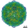
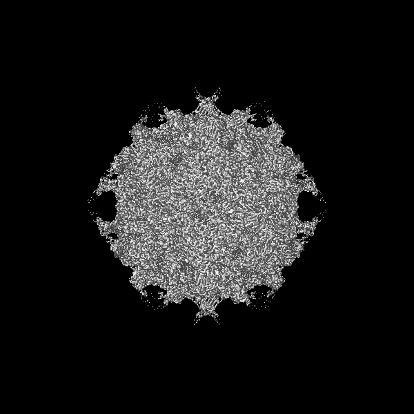
| Images | 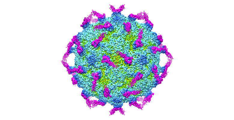 emd_30813.png emd_30813.png | 283.6 KB | ||
| Filedesc metadata | emd-30813.cif.gz | 6.1 KB | ||
| Archive directory |  http://ftp.pdbj.org/pub/emdb/structures/EMD-30813ftp://ftp.pdbj.org/pub/emdb/structures/EMD-30813 http://ftp.pdbj.org/pub/emdb/structures/EMD-30813ftp://ftp.pdbj.org/pub/emdb/structures/EMD-30813 | HTTPS FTP |
-Related structure data
| Related structure data |  7dq1MC  7dpfC  7dpgC  7dpzC  7dq4C  7dq7C M: atomic model generated by this map C: citing same article ( |
|---|---|
| Similar structure data |


-Links
| EMDB pages | EMDB (EBI/PDBe) / EMDataResource |
|---|---|
| Related items in Molecule of the Month |


-Map
| File | Download / File: emd_30813.map.gz / Format: CCP4 / Size: 824 MB / Type: IMAGE STORED AS FLOATING POINT NUMBER (4 BYTES) | ||||||||||||||||||||||||||||||||||||||||||||||||||||||||||||
|---|---|---|---|---|---|---|---|---|---|---|---|---|---|---|---|---|---|---|---|---|---|---|---|---|---|---|---|---|---|---|---|---|---|---|---|---|---|---|---|---|---|---|---|---|---|---|---|---|---|---|---|---|---|---|---|---|---|---|---|---|---|
| Annotation | Cryo-EM structure of CAR triggered Coxsachievirus B1 A-particle | ||||||||||||||||||||||||||||||||||||||||||||||||||||||||||||
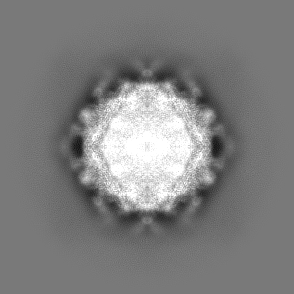
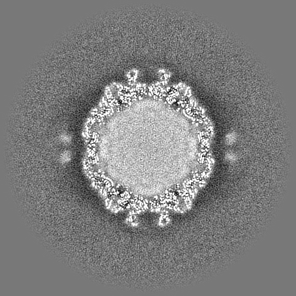
| Projections & slices | Image control
Images are generated by Spider. | ||||||||||||||||||||||||||||||||||||||||||||||||||||||||||||
| Voxel size | X=Y=Z: 1.12 Å | ||||||||||||||||||||||||||||||||||||||||||||||||||||||||||||
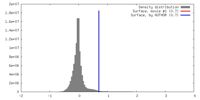
| Density |
| ||||||||||||||||||||||||||||||||||||||||||||||||||||||||||||
| Symmetry | Space group: 1 | ||||||||||||||||||||||||||||||||||||||||||||||||||||||||||||
| Details | EMDB XML:
CCP4 map header:
| ||||||||||||||||||||||||||||||||||||||||||||||||||||||||||||
 Z (Sec.)
 Y (Row.)
 X (Col.)


-Supplemental data
- Sample components
-Entire : Coxsackievirus B1
| Entire | Name: Coxsackievirus B1 |
|---|---|
| Components |
|
-Supramolecule #1: Coxsackievirus B1
| Supramolecule | Name: Coxsackievirus B1 / type: virus / ID: 1 / Parent: 0 / Macromolecule list: all / NCBI-ID: 12071 / Sci species name: Coxsackievirus B1 / Virus type: VIRION / Virus isolate: STRAIN / Virus enveloped: No / Virus empty: Yes |
|---|
-Macromolecule #1: Virion protein 1
| Macromolecule | Name: Virion protein 1 / type: protein_or_peptide / ID: 1 / Number of copies: 1 / Enantiomer: LEVO |
|---|---|
| Source (natural) | Organism: Coxsackievirus B1 |
| Molecular weight | Theoretical: 31.207117 KDa |
| Sequence | String: GPVEESVDRA VARVADTISS RPTNSESIPA LTAAETGHTS QVVPSDTMQT RHVKNYHSRS ESSIENFLCR SACVYYATYT NNSKKGFAE WVINTRQVAQ LRRKLELFTY LRFDLELTFV ITSAQQPSTA SSVDAPVQTH QIMYVPPGGP VPTKVKDYAW Q TSTNPSVF ...String: GPVEESVDRA VARVADTISS RPTNSESIPA LTAAETGHTS QVVPSDTMQT RHVKNYHSRS ESSIENFLCR SACVYYATYT NNSKKGFAE WVINTRQVAQ LRRKLELFTY LRFDLELTFV ITSAQQPSTA SSVDAPVQTH QIMYVPPGGP VPTKVKDYAW Q TSTNPSVF WTEGNAPPRM SIPFISIGNA YSCFYDGWTQ FSRNGVYGIN TLNNMGTLYM RHVNEAGQGP IKSTVRIYFK PK HVKAWVP RPPRLCQYEK QKNVNFSPIG VTTSRTDIIT T UniProtKB: Genome polyprotein |
-Macromolecule #2: VP2
| Macromolecule | Name: VP2 / type: protein_or_peptide / ID: 2 / Number of copies: 1 / Enantiomer: LEVO |
|---|---|
| Source (natural) | Organism: Coxsackievirus B1 |
| Molecular weight | Theoretical: 29.122744 KDa |
| Sequence | String: SPSAEECGYS DRVRSITLGN STITTQECAN VVVGYGVWPE YLKDNEATAE DQPTQPDVAT CRFYTLESVQ WMKNSAGWWW KLPDALSQM GLFGQNMQYH YLGRTGYTIH VQCNASKFHQ GCLLVVCVPE AEMGCSNLNN TPEFSELSGG DSARMFTDTQ V GESNAKKV ...String: SPSAEECGYS DRVRSITLGN STITTQECAN VVVGYGVWPE YLKDNEATAE DQPTQPDVAT CRFYTLESVQ WMKNSAGWWW KLPDALSQM GLFGQNMQYH YLGRTGYTIH VQCNASKFHQ GCLLVVCVPE AEMGCSNLNN TPEFSELSGG DSARMFTDTQ V GESNAKKV QTAVWNAGMG VGVGNLTIFP HQWINLRTNN SATLVMPYIN SVPMDNMFRH NNLTLMIIPF VPLNYSEGSS PY VPITVTI APMCAEYNGL RLASNQ UniProtKB: Genome polyprotein |
-Macromolecule #3: VP3
| Macromolecule | Name: VP3 / type: protein_or_peptide / ID: 3 / Number of copies: 1 / Enantiomer: LEVO |
|---|---|
| Source (natural) | Organism: Coxsackievirus B1 |
| Molecular weight | Theoretical: 26.328764 KDa |
| Sequence | String: GLPVMTTPGS TQFLTSDDFQ SPSAMPQFDV TPEMQIPGRV NNLMEIAEVD SVVPVNNTED NVSSLKAYQI PVQSNSDNGK QVFGFPLQP GANNVLNRTL LGEILNYYTH WSGSIKLTFM FCGSAMATGK FLLAYSPPGA GVPKNRKDAM LGTHVIWDVG L QSSCVLCV ...String: GLPVMTTPGS TQFLTSDDFQ SPSAMPQFDV TPEMQIPGRV NNLMEIAEVD SVVPVNNTED NVSSLKAYQI PVQSNSDNGK QVFGFPLQP GANNVLNRTL LGEILNYYTH WSGSIKLTFM FCGSAMATGK FLLAYSPPGA GVPKNRKDAM LGTHVIWDVG L QSSCVLCV PWISQTHYRY VVEDEYTAAG YVTCWYQTNI VVPADVQSSC DILCFVSACN DFSVRMLKDT PFIRQDTFYQ UniProtKB: Genome polyprotein |
-Macromolecule #4: Capsid protein VP4
| Macromolecule | Name: Capsid protein VP4 / type: protein_or_peptide / ID: 4 / Number of copies: 1 / Enantiomer: LEVO |
|---|---|
| Source (natural) | Organism: Coxsackievirus B1 |
| Molecular weight | Theoretical: 7.484246 KDa |
| Sequence | String: MGAQVSTQKT GAHETGLNAS GNSVIHYTNI NYYKDAASNS ANRQDFTQDP GKFTEPVKDI MVKTMPALN UniProtKB: Genome polyprotein |
-Macromolecule #5: Coxsackievirus and adenovirus receptor
| Macromolecule | Name: Coxsackievirus and adenovirus receptor / type: protein_or_peptide / ID: 5 / Number of copies: 1 / Enantiomer: LEVO |
|---|---|
| Source (natural) | Organism: Homo sapiens (human) |
| Molecular weight | Theoretical: 13.471361 KDa |
| Sequence | String: LSITTPEEMI EKAKGETAYL PCKFTLSPED QGPLDIEWLI SPADNQKVDQ VIILYSGDKI YDDYYPDLKG RVHFTSNDLK SGDASINVT NLQLSDIGTY QCKVKKAPGV ANKKIHLVVL VK UniProtKB: Coxsackievirus and adenovirus receptor |
-Experimental details
-Structure determination
| Method | cryo EM |
|---|---|
 Processing Processing | single particle reconstruction |
| Aggregation state | particle |
-Sample preparation
| Buffer | pH: 7.4 |
|---|---|
| Vitrification | Cryogen name: ETHANE |
- Electron microscopy
| Microscope | FEI TECNAI F30 |
|---|---|
| Image recording | Film or detector model: FEI FALCON III (4k x 4k) / Average electron dose: 25.0 e/Å2 |
| Electron beam | Acceleration voltage: 300 kV / Electron source:  FIELD EMISSION GUN FIELD EMISSION GUN |
| Electron optics | Illumination mode: FLOOD BEAM / Imaging mode: BRIGHT FIELD |
| Experimental equipment |  Model: Tecnai F30 / Image courtesy: FEI Company |
