 Movie
Movie Controller


+ Open data

- Basic information
| Entry | Database: PDB / ID: 1oop | ||||||
|---|---|---|---|---|---|---|---|
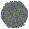
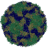
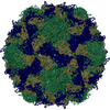
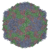
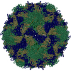
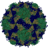
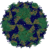
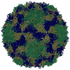
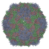
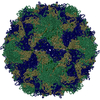
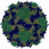
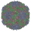
| Title | The Crystal Structure of Swine Vesicular Disease Virus | ||||||
 Components Components | (Coat protein ...) x 4 | ||||||
 Keywords Keywords | VIRUS / PICORNAVIRUS STRUCTURE / VIRUS/VIRAL PROTEIN / VIRUS-RECEPTOR INTERACTIONS / HOST ADAPTATION / CAR / DAF / COXSACKIEVIRUS / Icosahedral virus | ||||||
| Function / homology |  Function and homology information Function and homology informationsymbiont-mediated suppression of host cytoplasmic pattern recognition receptor signaling pathway via inhibition of RIG-I activity / picornain 2A / symbiont-mediated suppression of host mRNA export from nucleus / symbiont genome entry into host cell via pore formation in plasma membrane / picornain 3C / T=pseudo3 icosahedral viral capsid / host cell cytoplasmic vesicle membrane / ribonucleoside triphosphate phosphatase activity / nucleoside-triphosphate phosphatase / channel activity ...symbiont-mediated suppression of host cytoplasmic pattern recognition receptor signaling pathway via inhibition of RIG-I activity / picornain 2A / symbiont-mediated suppression of host mRNA export from nucleus / symbiont genome entry into host cell via pore formation in plasma membrane / picornain 3C / T=pseudo3 icosahedral viral capsid / host cell cytoplasmic vesicle membrane / ribonucleoside triphosphate phosphatase activity / nucleoside-triphosphate phosphatase / channel activity / monoatomic ion transmembrane transport / DNA replication / RNA helicase activity / endocytosis involved in viral entry into host cell / symbiont-mediated activation of host autophagy / RNA-directed RNA polymerase / cysteine-type endopeptidase activity / viral RNA genome replication / RNA-directed RNA polymerase activity / virion attachment to host cell / DNA-templated transcription / host cell nucleus / structural molecule activity / proteolysis / RNA binding / zinc ion binding / ATP binding Similarity search - Function | ||||||
| Biological species |  Swine vesicular disease virus Swine vesicular disease virus | ||||||
| Method |  X-RAY DIFFRACTION / SYNCHROTRON / MOLECULAR REPLACEMENT / Resolution: 3 Å X-RAY DIFFRACTION / SYNCHROTRON / MOLECULAR REPLACEMENT / Resolution: 3 Å | ||||||
 Authors Authors | Fry, E.E. / Knowles, N.J. / Newman, J.W.I. / Wilsden, G. / Rao, Z. / King, A.M.Q. / Stuart, D.I. | ||||||
 Citation Citation | Journal: J.Virol. / Year: 2003 Title: Crystal Structure of Swine Vesicular Disease Virus and Implications for Host Adaptation Authors: Fry, E.E. / Knowles, N.J. / Newman, J.W.I. / Wilsden, G. / Rao, Z. / King, A.M.Q. / Stuart, D.I. | ||||||
| History |
|
- Structure visualization
| Structure viewer | Molecule: MolmilJmol/JSmol |
|---|
- Downloads & links
-Download
| PDBx/mmCIF format | 1oop.cif.gz | 168.4 KB | Display | PDBx/mmCIF format |
|---|---|---|---|---|
| PDB format | pdb1oop.ent.gz | 131.9 KB | Display | PDB format |
| PDBx/mmJSON format | 1oop.json.gz | Tree view | PDBx/mmJSON format | |
| Others |  Other downloads Other downloads |
-Validation report
| Arichive directory | https://data.pdbj.org/pub/pdb/validation_reports/oo/1oopftp://data.pdbj.org/pub/pdb/validation_reports/oo/1oop | HTTPS FTP |
|---|
-Related structure data
| Similar structure data |
|---|


-Links
 PDBj
PDBj


- Assembly
| Deposited unit | 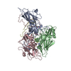
| ||||||||
|---|---|---|---|---|---|---|---|---|---|
| 1 | x 60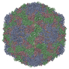
| ||||||||
| 2 |
| ||||||||
| 3 | x 5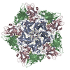
| ||||||||
| 4 | x 6
| ||||||||
| 5 | 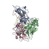
| ||||||||
| Unit cell |
| ||||||||
| Symmetry | Point symmetry: (Hermann–Mauguin notation: 532 / Schoenflies symbol: I (icosahedral)) |
-Components
-Coat protein ... , 4 types, 4 molecules ABCD
| #1: Protein | Mass: 31538.373 Da / Num. of mol.: 1 / Source method: isolated from a natural source Source: (natural) Swine vesicular disease virus (STRAIN UKG/27/72)Genus: Enterovirus / Species: Human enterovirus B / Strain: UKG-27-72 / References: UniProt: P13900 |
|---|---|
| #2: Protein | Mass: 28652.350 Da / Num. of mol.: 1 / Source method: isolated from a natural source Source: (natural) Swine vesicular disease virus (STRAIN UKG/27/72)Genus: Enterovirus / Species: Human enterovirus B / Strain: UKG-27-72 / References: UniProt: P13900 |
| #3: Protein | Mass: 26084.574 Da / Num. of mol.: 1 / Source method: isolated from a natural source Source: (natural) Swine vesicular disease virus (STRAIN UKG/27/72)Genus: Enterovirus / Species: Human enterovirus B / Strain: UKG-27-72 / References: UniProt: P13900 |
| #4: Protein | Mass: 7457.220 Da / Num. of mol.: 1 / Source method: isolated from a natural source Source: (natural) Swine vesicular disease virus (STRAIN UKG/27/72)Genus: Enterovirus / Species: Human enterovirus B / Strain: UKG-27-72 / References: UniProt: P13900 |
-Non-polymers , 2 types, 2 molecules 

| #5: Chemical | ChemComp-SPH /  Mass: 299.492 Da / Num. of mol.: 1 / Source method: obtained synthetically / Formula: C18H37NO2 Mass: 299.492 Da / Num. of mol.: 1 / Source method: obtained synthetically / Formula: C18H37NO2 |
|---|---|
| #6: Chemical | ChemComp-MYR /  Mass: 228.371 Da / Num. of mol.: 1 / Source method: obtained synthetically / Formula: C14H28O2 Mass: 228.371 Da / Num. of mol.: 1 / Source method: obtained synthetically / Formula: C14H28O2 |
-Experimental details
-Experiment
| Experiment | Method: X-RAY DIFFRACTION / Number of used crystals: 1 |
|---|
- Sample preparation
| Crystal grow | Temperature: 298 K / Method: vapor diffusion, sitting drop / pH: 7.6 Details: 15-25% saturated ammonium sulfate, 100mM phosphate buffer, pH 7.6, VAPOR DIFFUSION, SITTING DROP, temperature 298K | ||||||||||||||||||||||||
|---|---|---|---|---|---|---|---|---|---|---|---|---|---|---|---|---|---|---|---|---|---|---|---|---|---|
| Crystal grow | *PLUS | ||||||||||||||||||||||||
| Components of the solutions | *PLUS
|
-Data collection
| Diffraction | Mean temperature: 298 K |
|---|---|
| Diffraction source | Source: SYNCHROTRON / Site: SRS  / Beamline: PX14.2 / Wavelength: 0.979 Å / Beamline: PX14.2 / Wavelength: 0.979 Å |
| Detector | Type: ADSC QUANTUM 4 / Detector: CCD / Date: Feb 29, 2000 |
| Radiation | Protocol: SINGLE WAVELENGTH / Monochromatic (M) / Laue (L): M / Scattering type: x-ray |
| Radiation wavelength | Wavelength: 0.979 Å / Relative weight: 1 |
| Reflection | Resolution: 3→20 Å / Num. obs: 406689 / % possible obs: 49.2 % / Observed criterion σ(F): 0 / Observed criterion σ(I): 0 / Rmerge(I) obs: 0.207 / Net I/σ(I): 5.1 |
| Reflection shell | Resolution: 3→3.11 Å / Rmerge(I) obs: 0.482 / % possible all: 45.9 |
| Reflection shell | *PLUS % possible obs: 46 % |
- Processing
| Software |
| ||||||||||||||||||
|---|---|---|---|---|---|---|---|---|---|---|---|---|---|---|---|---|---|---|---|
| Refinement | Method to determine structure: MOLECULAR REPLACEMENT Starting model: Coxsackievirus A9 Resolution: 3→15 Å / σ(F): 0 / σ(I): 0 / Stereochemistry target values: Engh & Huber Details: A free R value is absent because the high non-crystallographic symmetry of viruses makes this less relevant.
| ||||||||||||||||||
| Displacement parameters | Biso mean: 14 Å2 | ||||||||||||||||||
| Refinement step | Cycle: LAST / Resolution: 3→15 Å
| ||||||||||||||||||
| Refine LS restraints |
| ||||||||||||||||||
| Refinement | *PLUS Highest resolution: 3 Å / Lowest resolution: 15 Å | ||||||||||||||||||
| Solvent computation | *PLUS | ||||||||||||||||||
| Displacement parameters | *PLUS |
