 Movie
Movie Controller


+ Open data

- Basic information
| Entry | Database: PDB / ID: 1mqt | ||||||
|---|---|---|---|---|---|---|---|
| Title | Swine Vesicular Disease Virus coat protein | ||||||
 Components Components |
| ||||||
 Keywords Keywords | VIRUS / swine vesicular disease virus / SVDV coat protein / enterovirus / Icosahedral virus | ||||||
| Function / homology |  Function and homology information Function and homology informationsymbiont genome entry into host cell via pore formation in plasma membrane / viral capsid / host cell cytoplasm / symbiont-mediated suppression of host gene expression / virion attachment to host cell / structural molecule activity Similarity search - Function | ||||||
| Biological species |  Swine vesicular disease virus Swine vesicular disease virus | ||||||
| Method |  X-RAY DIFFRACTION / SYNCHROTRON / MOLECULAR REPLACEMENT / Resolution: 3.3 Å X-RAY DIFFRACTION / SYNCHROTRON / MOLECULAR REPLACEMENT / Resolution: 3.3 Å | ||||||
 Authors Authors | Verdaguer, N. / Jimenez-Clavero, M.A. / Fita, I. / Ley, V. | ||||||
 Citation Citation | Journal: J.Virol. / Year: 2003 Title: STRUCTURE OF SWINE VESICULAR DISEASE VIRUS: MAPPING OF CHANGES OCCURRING DURING ADAPTATION OF HUMAN COXSACKIE B5 VIRUS TO INFECT SWINE Authors: Verdaguer, N. / Jimenez-Clavero, M.A. / Fita, I. / Ley, V. | ||||||
| History |
| ||||||
| Remark 600 | HETEROGEN The first residue of chain D is a myristic acid covalently attached to GLY D2 (not ...HETEROGEN The first residue of chain D is a myristic acid covalently attached to GLY D2 (not visible in the electron density) |
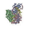
- Structure visualization
| Structure viewer | Molecule: MolmilJmol/JSmol |
|---|
- Downloads & links
-Download
| PDBx/mmCIF format | 1mqt.cif.gz | 170.2 KB | Display | PDBx/mmCIF format |
|---|---|---|---|---|
| PDB format | pdb1mqt.ent.gz | 134.3 KB | Display | PDB format |
| PDBx/mmJSON format | 1mqt.json.gz | Tree view | PDBx/mmJSON format | |
| Others |  Other downloads Other downloads |
-Validation report
| Arichive directory | https://data.pdbj.org/pub/pdb/validation_reports/mq/1mqtftp://data.pdbj.org/pub/pdb/validation_reports/mq/1mqt | HTTPS FTP |
|---|
-Related structure data
| Related structure data | 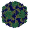 1covS S: Starting model for refinement |
|---|---|
| Similar structure data |


-Links
 PDBj
PDBj
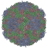
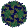
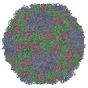
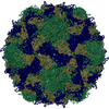
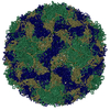
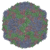
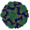
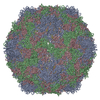
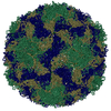
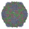
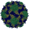
- Assembly
| Deposited unit | 
| ||||||||||||||||||||||||||||||||||||||||||||||||
|---|---|---|---|---|---|---|---|---|---|---|---|---|---|---|---|---|---|---|---|---|---|---|---|---|---|---|---|---|---|---|---|---|---|---|---|---|---|---|---|---|---|---|---|---|---|---|---|---|---|
| 1 | x 60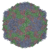
| ||||||||||||||||||||||||||||||||||||||||||||||||
| 2 |
| ||||||||||||||||||||||||||||||||||||||||||||||||
| 3 | x 5
| ||||||||||||||||||||||||||||||||||||||||||||||||
| 4 | x 6
| ||||||||||||||||||||||||||||||||||||||||||||||||
| 5 | 
| ||||||||||||||||||||||||||||||||||||||||||||||||
| 6 | x 15
| ||||||||||||||||||||||||||||||||||||||||||||||||
| Unit cell |
| ||||||||||||||||||||||||||||||||||||||||||||||||
| Symmetry | Point symmetry: (Hermann–Mauguin notation: 532 / Schoenflies symbol: I (icosahedral)) | ||||||||||||||||||||||||||||||||||||||||||||||||
| Noncrystallographic symmetry (NCS) | NCS oper:
|
-Components
-Protein , 1 types, 1 molecules A
| #1: Protein | Mass: 31370.240 Da / Num. of mol.: 1 / Fragment: SVDV COAT PROTEIN VP1 / Source method: isolated from a natural source / Source: (natural) Swine vesicular disease virus / Genus: Enterovirus / Species: Human enterovirus B / Strain: ISOLATE (SPA-2-'93) / References: UniProt: Q8B8X4 |
|---|
-Polyprotein Capsid ... , 3 types, 3 molecules BCD
| #2: Protein | Mass: 28505.158 Da / Num. of mol.: 1 / Fragment: SVDV COAT PROTEIN VP2 / Source method: isolated from a natural source / Source: (natural) Swine vesicular disease virus / Genus: Enterovirus / Species: Human enterovirus B / Strain: ISOLATE (SPA-2-'93) / References: UniProt: Q8B8X4 |
|---|---|
| #3: Protein | Mass: 26071.531 Da / Num. of mol.: 1 / Fragment: SVDV COAT PROTEIN VP3 / Source method: isolated from a natural source / Source: (natural) Swine vesicular disease virus / Genus: Enterovirus / Species: Human enterovirus B / Strain: ISOLATE (SPA-2-'93) / References: UniProt: Q8B8X4 |
| #4: Protein | Mass: 7353.049 Da / Num. of mol.: 1 / Fragment: SVDV COAT PROTEIN VP4 / Source method: isolated from a natural source / Source: (natural) Swine vesicular disease virus / Genus: Enterovirus / Species: Human enterovirus B / Strain: ISOLATE (SPA-2-'93) / References: UniProt: Q8B8X4 |
-Non-polymers , 2 types, 31 molecules 

| #5: Chemical | ChemComp-SPL /  Mass: 425.688 Da / Num. of mol.: 1 / Source method: obtained synthetically / Formula: C26H51NO3 Mass: 425.688 Da / Num. of mol.: 1 / Source method: obtained synthetically / Formula: C26H51NO3 |
|---|---|
| #6: Water | ChemComp-HOH / Mass: 18.015 Da / Num. of mol.: 30 / Source method: isolated from a natural source / Formula: H2O |
-Experimental details
-Experiment
| Experiment | Method: X-RAY DIFFRACTION / Number of used crystals: 2 |
|---|
- Sample preparation
| Crystal grow | Temperature: 293 K / Method: vapor diffusion, hanging drop / pH: 6.5 Details: ammonium sulfate, Potassium phosphate, pH 6.5, VAPOR DIFFUSION, HANGING DROP, temperature 293K | ||||||||||||||||||||
|---|---|---|---|---|---|---|---|---|---|---|---|---|---|---|---|---|---|---|---|---|---|
| Crystal grow | *PLUS pH: 6.2 / Method: vapor diffusion, hanging dropDetails: Jimenez-Clavero, M.A., (2003) Acta Cryst., D59, 541. | ||||||||||||||||||||
| Components of the solutions | *PLUS
|
-Data collection
| Diffraction | Mean temperature: 100 K |
|---|---|
| Diffraction source | Source: SYNCHROTRON / Site: ESRF  / Beamline: ID14-4 / Wavelength: 0.939 Å / Beamline: ID14-4 / Wavelength: 0.939 Å |
| Detector | Type: ADSC QUANTUM 4 / Detector: CCD / Date: Jun 19, 2002 |
| Radiation | Protocol: SINGLE WAVELENGTH / Monochromatic (M) / Laue (L): M / Scattering type: x-ray |
| Radiation wavelength | Wavelength: 0.939 Å / Relative weight: 1 |
| Reflection | Resolution: 3.3→30 Å / Num. all: 214790 / Num. obs: 214790 / % possible obs: 60 % / Observed criterion σ(F): 1 / Observed criterion σ(I): 1 / Redundancy: 3.1 % / Biso Wilson estimate: 16.6 Å2 / Rmerge(I) obs: 0.196 / Net I/σ(I): 4.6 |
| Reflection shell | Resolution: 3.3→3.5 Å / Rmerge(I) obs: 0.27 / % possible all: 44.1 |
| Reflection | *PLUS Highest resolution: 3 Å / Num. obs: 288614 / % possible obs: 70 % / Num. measured all: 459326 / Rmerge(I) obs: 0.1 |
| Reflection shell | *PLUS Highest resolution: 3 Å / Lowest resolution: 3.1 Å / % possible obs: 51 % / Rmerge(I) obs: 0.191 |
- Processing
| Software |
| |||||||||||||||||||||||||
|---|---|---|---|---|---|---|---|---|---|---|---|---|---|---|---|---|---|---|---|---|---|---|---|---|---|---|
| Refinement | Method to determine structure: MOLECULAR REPLACEMENT Starting model: PDB ENTRY 1COV Resolution: 3.3→30 Å / Isotropic thermal model: Isotropic / Cross valid method: THROUGHOUT / σ(F): 2 / Stereochemistry target values: Engh & Huber
| |||||||||||||||||||||||||
| Displacement parameters | Biso mean: 11.97 Å2 | |||||||||||||||||||||||||
| Refinement step | Cycle: LAST / Resolution: 3.3→30 Å
| |||||||||||||||||||||||||
| Refine LS restraints |
| |||||||||||||||||||||||||
| Refinement | *PLUS Highest resolution: 3 Å / Lowest resolution: 30 Å / Num. reflection obs: 266789 / Rfactor Rwork: 0.235 | |||||||||||||||||||||||||
| Solvent computation | *PLUS | |||||||||||||||||||||||||
| Displacement parameters | *PLUS | |||||||||||||||||||||||||
| Refine LS restraints | *PLUS
|
