Movie
Movie Controller

[English] 日本語
 Yorodumi
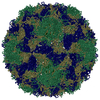
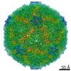
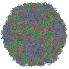
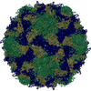
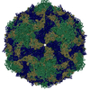
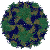
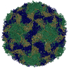
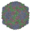
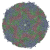
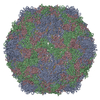
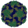
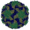
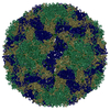
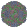
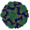
- PDB-6sk5: Cryo-EM structure of rhinovirus-B5 complexed to antiviral OBR-5-340 -
+ Open data

- Basic information
| Entry | Database: PDB / ID: 6sk5 | ||||||
|---|---|---|---|---|---|---|---|
| Title | Cryo-EM structure of rhinovirus-B5 complexed to antiviral OBR-5-340 | ||||||
 Components Components |
| ||||||
 Keywords Keywords | VIRUS / RV-A89 / HRV-B5 / capsid protein | ||||||
| Function / homology |  Function and homology information Function and homology informationpicornain 2A / symbiont-mediated suppression of host mRNA export from nucleus / symbiont genome entry into host cell via pore formation in plasma membrane / picornain 3C / T=pseudo3 icosahedral viral capsid / host cell cytoplasmic vesicle membrane / viral capsid / ribonucleoside triphosphate phosphatase activity / host cell / nucleoside-triphosphate phosphatase ...picornain 2A / symbiont-mediated suppression of host mRNA export from nucleus / symbiont genome entry into host cell via pore formation in plasma membrane / picornain 3C / T=pseudo3 icosahedral viral capsid / host cell cytoplasmic vesicle membrane / viral capsid / ribonucleoside triphosphate phosphatase activity / host cell / nucleoside-triphosphate phosphatase / channel activity / monoatomic ion transmembrane transport / RNA helicase activity / symbiont-mediated suppression of host innate immune response / endocytosis involved in viral entry into host cell / symbiont-mediated activation of host autophagy / RNA-directed RNA polymerase / cysteine-type endopeptidase activity / viral RNA genome replication / RNA-directed RNA polymerase activity / symbiont entry into host cell / virion attachment to host cell / host cell nucleus / structural molecule activity / DNA-templated transcription / proteolysis / RNA binding / zinc ion binding / ATP binding Similarity search - Function | ||||||
| Biological species |  Human rhinovirus B5 Human rhinovirus B5 | ||||||
| Method | ELECTRON MICROSCOPY / single particle reconstruction / cryo EM / Resolution: 3.6 Å | ||||||
 Authors Authors | Wald, J. / Goessweiner-Mohr, N. / Blaas, D. / Pasin, M. | ||||||
 Citation Citation | Journal: Proc Natl Acad Sci U S A / Year: 2019 Title: Cryo-EM structure of pleconaril-resistant rhinovirus-B5 complexed to the antiviral OBR-5-340 reveals unexpected binding site. Authors: Jiri Wald / Marion Pasin / Martina Richter / Christin Walther / Neann Mathai / Johannes Kirchmair / Vadim A Makarov / Nikolaus Goessweiner-Mohr / Thomas C Marlovits / Irene Zanella / Antonio ...Authors: Jiri Wald / Marion Pasin / Martina Richter / Christin Walther / Neann Mathai / Johannes Kirchmair / Vadim A Makarov / Nikolaus Goessweiner-Mohr / Thomas C Marlovits / Irene Zanella / Antonio Real-Hohn / Nuria Verdaguer / Dieter Blaas / Michaela Schmidtke /      Abstract: Viral inhibitors, such as pleconaril and vapendavir, target conserved regions in the capsids of rhinoviruses (RVs) and enteroviruses (EVs) by binding to a hydrophobic pocket in viral capsid protein 1 ...Viral inhibitors, such as pleconaril and vapendavir, target conserved regions in the capsids of rhinoviruses (RVs) and enteroviruses (EVs) by binding to a hydrophobic pocket in viral capsid protein 1 (VP1). In resistant RVs and EVs, bulky residues in this pocket prevent their binding. However, recently developed pyrazolopyrimidines inhibit pleconaril-resistant RVs and EVs, and computational modeling has suggested that they also bind to the hydrophobic pocket in VP1. We studied the mechanism of inhibition of pleconaril-resistant RVs using RV-B5 (1 of the 7 naturally pleconaril-resistant rhinoviruses) and OBR-5-340, a bioavailable pyrazolopyrimidine with proven in vivo activity, and determined the 3D-structure of the protein-ligand complex to 3.6 Å with cryoelectron microscopy. Our data indicate that, similar to other capsid binders, OBR-5-340 induces thermostability and inhibits viral adsorption and uncoating. However, we found that OBR-5-340 attaches closer to the entrance of the pocket than most other capsid binders, whose viral complexes have been studied so far, showing only marginal overlaps of the attachment sites. Comparing the experimentally determined 3D structure with the control, RV-B5 incubated with solvent only and determined to 3.2 Å, revealed no gross conformational changes upon OBR-5-340 binding. The pocket of the naturally OBR-5-340-resistant RV-A89 likewise incubated with OBR-5-340 and solved to 2.9 Å was empty. Pyrazolopyrimidines have a rigid molecular scaffold and may thus be less affected by a loss of entropy upon binding. They interact with less-conserved regions than known capsid binders. Overall, pyrazolopyrimidines could be more suitable for the development of new, broadly active inhibitors. | ||||||
| History |
|
- Structure visualization
| Movie |
Movie viewer |
|---|---|
| Structure viewer | Molecule: MolmilJmol/JSmol |
- Downloads & links
-Download
| PDBx/mmCIF format | 6sk5.cif.gz | 290.3 KB | Display | PDBx/mmCIF format |
|---|---|---|---|---|
| PDB format | pdb6sk5.ent.gz | 239.4 KB | Display | PDB format |
| PDBx/mmJSON format | 6sk5.json.gz | Tree view | PDBx/mmJSON format | |
| Others |  Other downloads Other downloads |
-Validation report
| Arichive directory | https://data.pdbj.org/pub/pdb/validation_reports/sk/6sk5ftp://data.pdbj.org/pub/pdb/validation_reports/sk/6sk5 | HTTPS FTP |
|---|
-Related structure data
| Related structure data |  10220MC  6sk6C  6sk7C M: map data used to model this data C: citing same article ( |
|---|---|
| Similar structure data |


-Links
 PDBj
PDBj

- Assembly
| Deposited unit | 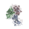
|
|---|---|
| 1 | x 60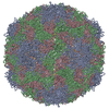
|
-Components
| #1: Protein | Mass: 7335.049 Da / Num. of mol.: 1 / Source method: isolated from a natural source / Details: Virus grown in human cell line / Source: (natural) Human rhinovirus B5 / References: UniProt: Q80SQ3 |
|---|---|
| #2: Protein | Mass: 27507.256 Da / Num. of mol.: 1 / Source method: isolated from a natural source / Details: Virus grown in human cell line / Source: (natural) Human rhinovirus B5References: UniProt: B9V433, picornain 2A, nucleoside-triphosphate phosphatase, picornain 3C, RNA-directed RNA polymerase |
| #3: Protein | Mass: 32743.861 Da / Num. of mol.: 1 / Source method: isolated from a natural source / Details: Virus grown in human cell line / Source: (natural) Human rhinovirus B5 / References: UniProt: Q7T659 |
| #4: Protein | Mass: 25500.168 Da / Num. of mol.: 1 / Source method: isolated from a natural source / Details: Virus grown in human cell line / Source: (natural) Human rhinovirus B5References: UniProt: B9V433, picornain 2A, nucleoside-triphosphate phosphatase, picornain 3C, RNA-directed RNA polymerase |
| #5: Chemical | ChemComp-LGQ / 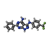  Mass: 370.331 Da / Num. of mol.: 1 / Source method: obtained synthetically / Formula: C18H13F3N6 / Feature type: SUBJECT OF INVESTIGATION Mass: 370.331 Da / Num. of mol.: 1 / Source method: obtained synthetically / Formula: C18H13F3N6 / Feature type: SUBJECT OF INVESTIGATION |
| Has ligand of interest | Y |
-Experimental details
-Experiment
| Experiment | Method: ELECTRON MICROSCOPY |
|---|---|
| EM experiment | Aggregation state: PARTICLE / 3D reconstruction method: single particle reconstruction |
- Sample preparation
| Component | Name: Human rhinovirus B5 / Type: VIRUS / Entity ID: #1-#4 / Source: NATURAL | ||||||||||||
|---|---|---|---|---|---|---|---|---|---|---|---|---|---|
| Molecular weight | Experimental value: NO | ||||||||||||
| Source (natural) | Organism: Human rhinovirus B5 / Strain: B5 | ||||||||||||
| Details of virus | Empty: NO / Enveloped: NO / Isolate: SEROTYPE / Type: VIRION | ||||||||||||
| Natural host | Organism: Homo sapiens | ||||||||||||
| Virus shell | Name: VP1-4 / Diameter: 302 nm | ||||||||||||
| Buffer solution | pH: 7.4 | ||||||||||||
| Buffer component |
| ||||||||||||
| Specimen | Embedding applied: NO / Shadowing applied: NO / Staining applied: NO / Vitrification applied: YES | ||||||||||||
| Specimen support | Grid material: COPPER / Grid type: Homemade | ||||||||||||
| Vitrification | Instrument: FEI VITROBOT MARK III / Cryogen name: ETHANE / Humidity: 100 % |
- Electron microscopy imaging
| Experimental equipment |  Model: Tecnai Polara / Image courtesy: FEI Company |
|---|---|
| Microscopy | Model: FEI POLARA 300 |
| Electron gun | Electron source:  FIELD EMISSION GUN / Accelerating voltage: 300 kV / Illumination mode: FLOOD BEAM FIELD EMISSION GUN / Accelerating voltage: 300 kV / Illumination mode: FLOOD BEAM |
| Electron lens | Mode: BRIGHT FIELD / Cs: 2.7 mm |
| Image recording | Average exposure time: 5 sec. / Electron dose: 40 e/Å2 / Detector mode: COUNTING / Film or detector model: GATAN K2 SUMMIT (4k x 4k) / Num. of real images: 2547 |
| Image scans | Movie frames/image: 25 / Used frames/image: 1-25 |
- Processing
| EM software |
| ||||||||||||||||||||
|---|---|---|---|---|---|---|---|---|---|---|---|---|---|---|---|---|---|---|---|---|---|
| CTF correction | Type: NONE | ||||||||||||||||||||
| Symmetry | Point symmetry: C60 (60 fold cyclic) | ||||||||||||||||||||
| 3D reconstruction | Resolution: 3.6 Å / Resolution method: FSC 0.143 CUT-OFF / Num. of particles: 46070 / Symmetry type: POINT | ||||||||||||||||||||
| Atomic model building | Space: REAL Details: Rosetta: DiMaio, F. et al. Atomic-accuracy models from 4.5-A cryo-electron microscopy data with density-guided iterative local refinement. Nat. Meth (2015). doi:10.1038/nmeth.3286 | ||||||||||||||||||||
| Atomic model building | PDB-ID: 1AYM Accession code: 1AYM / Source name: PDB / Type: experimental model |