 Movie
Movie Controller


+ Open data

- Basic information
| Entry | Database: PDB / ID: 4gb3 | ||||||
|---|---|---|---|---|---|---|---|
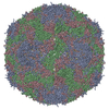
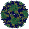
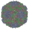
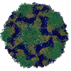
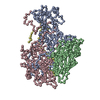
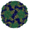
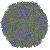
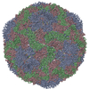
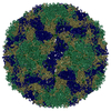
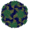
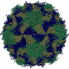
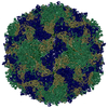
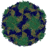
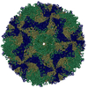
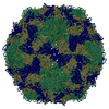
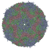
| Title | Human coxsackievirus B3 strain RD coat protein | ||||||
 Components Components | (coat protein ...) x 4 | ||||||
 Keywords Keywords | VIRUS / Capsid Protein | ||||||
| Function / homology |  Function and homology information Function and homology informationsymbiont-mediated suppression of host cytoplasmic pattern recognition receptor signaling pathway via inhibition of RIG-I activity / picornain 2A / symbiont-mediated suppression of host mRNA export from nucleus / symbiont genome entry into host cell via pore formation in plasma membrane / picornain 3C / T=pseudo3 icosahedral viral capsid / host cell cytoplasmic vesicle membrane / ribonucleoside triphosphate phosphatase activity / nucleoside-triphosphate phosphatase / channel activity ...symbiont-mediated suppression of host cytoplasmic pattern recognition receptor signaling pathway via inhibition of RIG-I activity / picornain 2A / symbiont-mediated suppression of host mRNA export from nucleus / symbiont genome entry into host cell via pore formation in plasma membrane / picornain 3C / T=pseudo3 icosahedral viral capsid / host cell cytoplasmic vesicle membrane / ribonucleoside triphosphate phosphatase activity / nucleoside-triphosphate phosphatase / channel activity / monoatomic ion transmembrane transport / DNA replication / RNA helicase activity / endocytosis involved in viral entry into host cell / symbiont-mediated activation of host autophagy / RNA-directed RNA polymerase / cysteine-type endopeptidase activity / viral RNA genome replication / RNA-directed RNA polymerase activity / virion attachment to host cell / host cell nucleus / structural molecule activity / DNA-templated transcription / proteolysis / RNA binding / zinc ion binding / ATP binding Similarity search - Function | ||||||
| Biological species |   Human coxsackievirus B3 Human coxsackievirus B3 | ||||||
| Method |  X-RAY DIFFRACTION / SYNCHROTRON / MOLECULAR REPLACEMENT / Resolution: 2.74 Å X-RAY DIFFRACTION / SYNCHROTRON / MOLECULAR REPLACEMENT / Resolution: 2.74 Å | ||||||
 Authors Authors | Yoder, J.D. / Hafenstein, S. | ||||||
 Citation Citation | Journal: J Virol / Year: 2012 Title: The crystal structure of a coxsackievirus B3-RD variant and a refined 9-angstrom cryo-electron microscopy reconstruction of the virus complexed with decay-accelerating factor (DAF) provide a ...Title: The crystal structure of a coxsackievirus B3-RD variant and a refined 9-angstrom cryo-electron microscopy reconstruction of the virus complexed with decay-accelerating factor (DAF) provide a new footprint of DAF on the virus surface. Authors: Joshua D Yoder / Javier O Cifuente / Jieyan Pan / Jeffrey M Bergelson / Susan Hafenstein /  Abstract: The coxsackievirus-adenovirus receptor (CAR) and decay-accelerating factor (DAF) have been identified as cellular receptors for coxsackievirus B3 (CVB3). The first described DAF-binding isolate was ...The coxsackievirus-adenovirus receptor (CAR) and decay-accelerating factor (DAF) have been identified as cellular receptors for coxsackievirus B3 (CVB3). The first described DAF-binding isolate was obtained during passage of the prototype strain, Nancy, on rhabdomyosarcoma (RD) cells, which express DAF but very little CAR. Here, the structure of the resulting variant, CVB3-RD, has been solved by X-ray crystallography to 2.74 Å, and a cryo-electron microscopy reconstruction of CVB3-RD complexed with DAF has been refined to 9.0 Å. This new high-resolution structure permits us to correct an error in our previous view of DAF-virus interactions, providing a new footprint of DAF that bridges two adjacent protomers. The contact sites between the virus and DAF clearly encompass CVB3-RD residues recently shown to be required for binding to DAF; these residues interact with DAF short consensus repeat 2 (SCR2), which is known to be essential for virus binding. Based on the new structure, the mode of the DAF interaction with CVB3 differs significantly from the mode reported previously for DAF binding to echoviruses. | ||||||
| History |
|
- Structure visualization
| Structure viewer | Molecule: MolmilJmol/JSmol |
|---|
- Downloads & links
-Download
| PDBx/mmCIF format | 4gb3.cif.gz | 174.1 KB | Display | PDBx/mmCIF format |
|---|---|---|---|---|
| PDB format | pdb4gb3.ent.gz | 136.5 KB | Display | PDB format |
| PDBx/mmJSON format | 4gb3.json.gz | Tree view | PDBx/mmJSON format | |
| Others |  Other downloads Other downloads |
-Validation report
| Arichive directory | https://data.pdbj.org/pub/pdb/validation_reports/gb/4gb3ftp://data.pdbj.org/pub/pdb/validation_reports/gb/4gb3 | HTTPS FTP |
|---|
-Related structure data
| Related structure data | 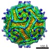 5475C  3j24C 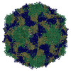 1covS S: Starting model for refinement C: citing same article ( |
|---|---|
| Similar structure data |


-Links
 PDBj
PDBj


- Assembly
| Deposited unit | 
| ||||||||||||||||||||||||||||||||||||||||||||
|---|---|---|---|---|---|---|---|---|---|---|---|---|---|---|---|---|---|---|---|---|---|---|---|---|---|---|---|---|---|---|---|---|---|---|---|---|---|---|---|---|---|---|---|---|---|
| 1 | x 60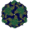
| ||||||||||||||||||||||||||||||||||||||||||||
| 2 |
| ||||||||||||||||||||||||||||||||||||||||||||
| 3 | x 5
| ||||||||||||||||||||||||||||||||||||||||||||
| 4 | x 6
| ||||||||||||||||||||||||||||||||||||||||||||
| 5 | 
| ||||||||||||||||||||||||||||||||||||||||||||
| 6 | x 10
| ||||||||||||||||||||||||||||||||||||||||||||
| Unit cell |
| ||||||||||||||||||||||||||||||||||||||||||||
| Symmetry | Point symmetry: (Schoenflies symbol: I (icosahedral)) | ||||||||||||||||||||||||||||||||||||||||||||
| Noncrystallographic symmetry (NCS) | NCS oper:
|
-Components
-Coat protein ... , 4 types, 4 molecules 1234
| #1: Protein | Mass: 31364.068 Da / Num. of mol.: 1 / Fragment: UNP residues 571-851 / Source method: isolated from a natural source / Source: (natural) Human coxsackievirus B3 / Strain: RD / References: UniProt: F8VA14 |
|---|---|
| #2: Protein | Mass: 28878.580 Da / Num. of mol.: 1 / Fragment: UNP residues 70-332 / Source method: isolated from a natural source / Source: (natural) Human coxsackievirus B3 / Strain: RD / References: UniProt: F8VA14 |
| #3: Protein | Mass: 26211.725 Da / Num. of mol.: 1 / Fragment: UNP residues 333-570 / Source method: isolated from a natural source / Source: (natural) Human coxsackievirus B3 / Strain: RD / References: UniProt: F8VA14 |
| #4: Protein | Mass: 7349.039 Da / Num. of mol.: 1 / Fragment: UNP residues 2-69 / Source method: isolated from a natural source / Source: (natural) Human coxsackievirus B3 / Strain: RD / References: UniProt: F8VA14 |
-Non-polymers , 2 types, 2 molecules 

| #5: Chemical | ChemComp-PLM /  Mass: 256.424 Da / Num. of mol.: 1 / Source method: obtained synthetically / Formula: C16H32O2 Mass: 256.424 Da / Num. of mol.: 1 / Source method: obtained synthetically / Formula: C16H32O2 |
|---|---|
| #6: Chemical | ChemComp-MYR /  Mass: 228.371 Da / Num. of mol.: 1 / Source method: obtained synthetically / Formula: C14H28O2 Mass: 228.371 Da / Num. of mol.: 1 / Source method: obtained synthetically / Formula: C14H28O2 |
-Details
| Has protein modification | Y |
|---|
-Experimental details
-Experiment
| Experiment | Method: X-RAY DIFFRACTION / Number of used crystals: 1 |
|---|
- Sample preparation
| Crystal | Density Matthews: 3.67 Å3/Da / Density % sol: 66.49 % |
|---|---|
| Crystal grow | Temperature: 298 K / pH: 6 Details: 2 M ammonium sulfate, pH 6.0, VAPOR DIFFUSION, SITTING DROP, temperature 298.0K |
-Data collection
| Diffraction | Mean temperature: 100 K |
|---|---|
| Diffraction source | Source: SYNCHROTRON / Site: APS / Beamline: 14-BM-C / Wavelength: 0.979 |
| Detector | Type: ADSC QUANTUM 315 / Detector: CCD / Date: Feb 7, 2008 |
| Radiation | Monochromator: BENT GE(111) / Protocol: SINGLE WAVELENGTH / Monochromatic (M) / Laue (L): M / Scattering type: x-ray |
| Radiation wavelength | Wavelength: 0.979 Å / Relative weight: 1 |
| Reflection | Resolution: 2.74→15 Å / Num. obs: 343901 / % possible obs: 96.9 % / Observed criterion σ(I): 2 / Redundancy: 4.4 % / Biso Wilson estimate: 40.5 Å2 / Rmerge(I) obs: 0.127 / Net I/σ(I): 8.75 |
| Reflection shell | Resolution: 2.74→2.84 Å / Redundancy: 4.4 % / Rmerge(I) obs: 0.488 / Mean I/σ(I) obs: 2.97 / % possible all: 98 |
- Processing
| Software |
| ||||||||||||||||||||||||||||||||||||||||||||||||||||||||||||
|---|---|---|---|---|---|---|---|---|---|---|---|---|---|---|---|---|---|---|---|---|---|---|---|---|---|---|---|---|---|---|---|---|---|---|---|---|---|---|---|---|---|---|---|---|---|---|---|---|---|---|---|---|---|---|---|---|---|---|---|---|---|
| Refinement | Method to determine structure: MOLECULAR REPLACEMENT Starting model: PDB ENTRY 1COV Resolution: 2.74→15 Å / Rfactor Rfree error: 0.003 / Data cutoff high absF: 72829.27 / Data cutoff low absF: 0 / Cross valid method: THROUGHOUT / σ(F): 0 / Stereochemistry target values: ENGH & HUBER
| ||||||||||||||||||||||||||||||||||||||||||||||||||||||||||||
| Solvent computation | Solvent model: FLAT MODEL / Bsol: 18.1026 Å2 / ksol: 0.34 e/Å3 | ||||||||||||||||||||||||||||||||||||||||||||||||||||||||||||
| Displacement parameters | Biso mean: 44.8 Å2
| ||||||||||||||||||||||||||||||||||||||||||||||||||||||||||||
| Refine analyze |
| ||||||||||||||||||||||||||||||||||||||||||||||||||||||||||||
| Refinement step | Cycle: LAST / Resolution: 2.74→15 Å
| ||||||||||||||||||||||||||||||||||||||||||||||||||||||||||||
| Refine LS restraints |
| ||||||||||||||||||||||||||||||||||||||||||||||||||||||||||||
| LS refinement shell | Resolution: 2.74→2.91 Å / Rfactor Rfree error: 0.009 / Total num. of bins used: 6
|
