Movie
Movie Controller

[English] 日本語
 Yorodumi
- EMDB-5475: Cryo-EM reconstruction of Coxsackievirus B3 strain RD complexed w... -
+ Open data

- Basic information
| Entry | Database: EMDB / ID: EMD-5475 | |||||||||
|---|---|---|---|---|---|---|---|---|---|---|
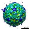
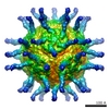
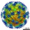
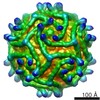
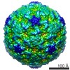
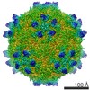
| Title | Cryo-EM reconstruction of Coxsackievirus B3 strain RD complexed with receptor DAF | |||||||||
 Map data Map data | Cryo-electron microscopy reconstruction of Coxsackievirus B3 strain RD complexed with decay accelerating factor SCR1-4 | |||||||||
 Sample Sample |
| |||||||||
 Keywords Keywords | coxsackievirus / enterovirus / picornavirus / receptor / DAF | |||||||||
| Function / homology |  Function and homology information Function and homology informationregulation of lipopolysaccharide-mediated signaling pathway / negative regulation of complement activation / regulation of complement-dependent cytotoxicity / regulation of complement activation / respiratory burst / positive regulation of CD4-positive, alpha-beta T cell activation / positive regulation of CD4-positive, alpha-beta T cell proliferation / Class B/2 (Secretin family receptors) / ficolin-1-rich granule membrane / complement activation, classical pathway ...regulation of lipopolysaccharide-mediated signaling pathway / negative regulation of complement activation / regulation of complement-dependent cytotoxicity / regulation of complement activation / respiratory burst / positive regulation of CD4-positive, alpha-beta T cell activation / positive regulation of CD4-positive, alpha-beta T cell proliferation / Class B/2 (Secretin family receptors) / ficolin-1-rich granule membrane / complement activation, classical pathway / side of membrane / transport vesicle / COPI-mediated anterograde transport / endoplasmic reticulum-Golgi intermediate compartment membrane / Regulation of Complement cascade / secretory granule membrane / positive regulation of T cell cytokine production / virus receptor activity / positive regulation of cytosolic calcium ion concentration / membrane raft / Golgi membrane / innate immune response / Neutrophil degranulation / lipid binding / cell surface / extracellular exosome / extracellular region / plasma membrane Similarity search - Function | |||||||||
| Biological species |  Homo sapiens (human) / Homo sapiens (human) /   Human coxsackievirus B3 Human coxsackievirus B3 | |||||||||
| Method | single particle reconstruction / cryo EM / Resolution: 9.0 Å | |||||||||
 Authors Authors | Yoder JD / Cifuente JO / Pan J / Bergelson JM / Hafenstein S | |||||||||
 Citation Citation | Journal: J Virol / Year: 2012 Title: The crystal structure of a coxsackievirus B3-RD variant and a refined 9-angstrom cryo-electron microscopy reconstruction of the virus complexed with decay-accelerating factor (DAF) provide a ...Title: The crystal structure of a coxsackievirus B3-RD variant and a refined 9-angstrom cryo-electron microscopy reconstruction of the virus complexed with decay-accelerating factor (DAF) provide a new footprint of DAF on the virus surface. Authors: Joshua D Yoder / Javier O Cifuente / Jieyan Pan / Jeffrey M Bergelson / Susan Hafenstein /  Abstract: The coxsackievirus-adenovirus receptor (CAR) and decay-accelerating factor (DAF) have been identified as cellular receptors for coxsackievirus B3 (CVB3). The first described DAF-binding isolate was ...The coxsackievirus-adenovirus receptor (CAR) and decay-accelerating factor (DAF) have been identified as cellular receptors for coxsackievirus B3 (CVB3). The first described DAF-binding isolate was obtained during passage of the prototype strain, Nancy, on rhabdomyosarcoma (RD) cells, which express DAF but very little CAR. Here, the structure of the resulting variant, CVB3-RD, has been solved by X-ray crystallography to 2.74 Å, and a cryo-electron microscopy reconstruction of CVB3-RD complexed with DAF has been refined to 9.0 Å. This new high-resolution structure permits us to correct an error in our previous view of DAF-virus interactions, providing a new footprint of DAF that bridges two adjacent protomers. The contact sites between the virus and DAF clearly encompass CVB3-RD residues recently shown to be required for binding to DAF; these residues interact with DAF short consensus repeat 2 (SCR2), which is known to be essential for virus binding. Based on the new structure, the mode of the DAF interaction with CVB3 differs significantly from the mode reported previously for DAF binding to echoviruses. | |||||||||
| History |
|
- Structure visualization
| Movie |
Movie viewer |
|---|---|
| Structure viewer | EM map: SurfViewMolmilJmol/JSmol |
| Supplemental images |
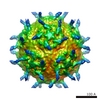
 UCSF Chimera
- Downloads & links
-EMDB archive
| Map data | emd_5475.map.gz | 7.1 MB | EMDB map data format | |
|---|---|---|---|---|
| Header (meta data) | emd-5475-v30.xmlemd-5475.xml | 12.4 KB 12.4 KB | Display Display | EMDB header |
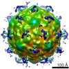
| Images | 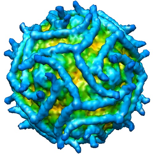 emd_5475_1.jpg emd_5475_1.jpg | 76.1 KB | ||
| Archive directory |  http://ftp.pdbj.org/pub/emdb/structures/EMD-5475ftp://ftp.pdbj.org/pub/emdb/structures/EMD-5475 http://ftp.pdbj.org/pub/emdb/structures/EMD-5475ftp://ftp.pdbj.org/pub/emdb/structures/EMD-5475 | HTTPS FTP |
-Related structure data
| Related structure data |  3j24MC  4gb3C M: atomic model generated by this map C: citing same article ( |
|---|---|
| Similar structure data |
-Links
| EMDB pages | EMDB (EBI/PDBe) / EMDataResource |
|---|---|
| Related items in Molecule of the Month |


-Map
| File | Download / File: emd_5475.map.gz / Format: CCP4 / Size: 18.6 MB / Type: IMAGE STORED AS FLOATING POINT NUMBER (4 BYTES) | ||||||||||||||||||||||||||||||||||||||||||||||||||||||||||||||||||||
|---|---|---|---|---|---|---|---|---|---|---|---|---|---|---|---|---|---|---|---|---|---|---|---|---|---|---|---|---|---|---|---|---|---|---|---|---|---|---|---|---|---|---|---|---|---|---|---|---|---|---|---|---|---|---|---|---|---|---|---|---|---|---|---|---|---|---|---|---|---|
| Annotation | Cryo-electron microscopy reconstruction of Coxsackievirus B3 strain RD complexed with decay accelerating factor SCR1-4 | ||||||||||||||||||||||||||||||||||||||||||||||||||||||||||||||||||||
| Projections & slices | Image control
Images are generated by Spider. | ||||||||||||||||||||||||||||||||||||||||||||||||||||||||||||||||||||
| Voxel size | X=Y=Z: 2.94 Å | ||||||||||||||||||||||||||||||||||||||||||||||||||||||||||||||||||||
| Density |
| ||||||||||||||||||||||||||||||||||||||||||||||||||||||||||||||||||||
| Symmetry | Space group: 1 | ||||||||||||||||||||||||||||||||||||||||||||||||||||||||||||||||||||
| Details | EMDB XML:
CCP4 map header:
| ||||||||||||||||||||||||||||||||||||||||||||||||||||||||||||||||||||
 Z (Sec.)
 Y (Row.)
 X (Col.)


-Supplemental data
- Sample components
-Entire : Coxsackievirus B3 strain RD (CVB3-RD), complexed with decay-accel...
| Entire | Name: Coxsackievirus B3 strain RD (CVB3-RD), complexed with decay-accelerating factor (DAF) |
|---|---|
| Components |
|
-Supramolecule #1000: Coxsackievirus B3 strain RD (CVB3-RD), complexed with decay-accel...
| Supramolecule | Name: Coxsackievirus B3 strain RD (CVB3-RD), complexed with decay-accelerating factor (DAF) type: sample / ID: 1000 Details: One DAF binds each binding site (one per CVB3-RD protomer). Oligomeric state: One receptor per virus protomer / Number unique components: 2 |
|---|---|
| Molecular weight | Theoretical: 7 MDa |
-Supramolecule #1: Human coxsackievirus B3
| Supramolecule | Name: Human coxsackievirus B3 / type: virus / ID: 1 / NCBI-ID: 12072 / Sci species name: Human coxsackievirus B3 / Database: NCBI / Virus type: VIRION / Virus isolate: STRAIN / Virus enveloped: No / Virus empty: No |
|---|---|
| Host (natural) | Organism: Homo sapiens (human) / synonym: VERTEBRATES |
| Molecular weight | Theoretical: 7 MDa |
| Virus shell | Shell ID: 1 / Diameter: 300 Å / T number (triangulation number): 1 |
-Macromolecule #1: decay-accelerating factor
| Macromolecule | Name: decay-accelerating factor / type: protein_or_peptide / ID: 1 / Name.synonym: DAF / Number of copies: 60 / Recombinant expression: Yes |
|---|---|
| Source (natural) | Organism: Homo sapiens (human) / synonym: human |
| Recombinant expression | Organism:  |
-Experimental details
-Structure determination
| Method | cryo EM |
|---|---|
 Processing Processing | single particle reconstruction |
| Aggregation state | particle |
-Sample preparation
| Concentration | 2.0 mg/mL |
|---|---|
| Buffer | pH: 6 / Details: 50mM MES |
| Grid | Details: Quantifoil |
| Vitrification | Cryogen name: ETHANE / Chamber temperature: 120 K / Instrument: HOMEMADE PLUNGER / Method: Blot before plunging |
- Electron microscopy
| Microscope | FEI/PHILIPS CM300FEG/T |
|---|---|
| Temperature | Min: 83 K / Max: 93 K |
| Alignment procedure | Legacy - Astigmatism: Lens astigmatism was corrected at 98,000 times magnification Legacy - Electron beam tilt params: 0 |
| Date | Aug 6, 2004 |
| Image recording | Category: FILM / Film or detector model: KODAK SO-163 FILM / Digitization - Scanner: ZEISS SCAI / Number real images: 36 / Average electron dose: 24 e/Å2 / Details: scanned at 7 microns and bin-averaged to 14 / Od range: 1 / Bits/pixel: 8 |
| Electron beam | Acceleration voltage: 300 kV / Electron source: TUNGSTEN HAIRPIN |
| Electron optics | Calibrated magnification: 47000 / Illumination mode: FLOOD BEAM / Imaging mode: BRIGHT FIELD / Cs: 2.0 mm / Nominal defocus max: 4.6 µm / Nominal defocus min: 1.0 µm / Nominal magnification: 45000 |
| Sample stage | Specimen holder: Side mounted nitrogen cooled / Specimen holder model: GATAN LIQUID NITROGEN |
-Image processing
| Details | Using the program Robem, particles were picked from 36 of the highest quality micrographs with a selection area sized to 171x171 pixels and preprocessed using program autopp to remove blemishes, linearize, normalize, and apodize. To correct for contrast transfer function, defocus and astigmatism values were assessed from the digitized images using the program ctffind3. |
|---|---|
| CTF correction | Details: AUTO3DEM |
| Final reconstruction | Algorithm: OTHER / Resolution.type: BY AUTHOR / Resolution: 9.0 Å / Resolution method: FSC 0.5 CUT-OFF / Software - Name: AUTO3DEM, CTFFIND, autopp, Robem / Number images used: 3010 |