 Movie
Movie Controller
 Structure viewers
 About Yorodumi Papers


+Search query
-Structure paper
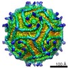
| Title | The crystal structure of a coxsackievirus B3-RD variant and a refined 9-angstrom cryo-electron microscopy reconstruction of the virus complexed with decay-accelerating factor (DAF) provide a new footprint of DAF on the virus surface. |
|---|---|
| Journal, issue, pages | J Virol, Vol. 86, Issue 23, Page 12571-12581, Year 2012 |
| Publish date | Sep 12, 2012 |
 Authors Authors | Joshua D Yoder / Javier O Cifuente / Jieyan Pan / Jeffrey M Bergelson / Susan Hafenstein /  |
| PubMed Abstract | The coxsackievirus-adenovirus receptor (CAR) and decay-accelerating factor (DAF) have been identified as cellular receptors for coxsackievirus B3 (CVB3). The first described DAF-binding isolate was ...The coxsackievirus-adenovirus receptor (CAR) and decay-accelerating factor (DAF) have been identified as cellular receptors for coxsackievirus B3 (CVB3). The first described DAF-binding isolate was obtained during passage of the prototype strain, Nancy, on rhabdomyosarcoma (RD) cells, which express DAF but very little CAR. Here, the structure of the resulting variant, CVB3-RD, has been solved by X-ray crystallography to 2.74 Å, and a cryo-electron microscopy reconstruction of CVB3-RD complexed with DAF has been refined to 9.0 Å. This new high-resolution structure permits us to correct an error in our previous view of DAF-virus interactions, providing a new footprint of DAF that bridges two adjacent protomers. The contact sites between the virus and DAF clearly encompass CVB3-RD residues recently shown to be required for binding to DAF; these residues interact with DAF short consensus repeat 2 (SCR2), which is known to be essential for virus binding. Based on the new structure, the mode of the DAF interaction with CVB3 differs significantly from the mode reported previously for DAF binding to echoviruses. |
 External links External links | J Virol / PubMed:22973031 / PubMed Central |
| Methods | EM (single particle) / X-ray diffraction |
| Resolution | 2.74 - 9.0 Å |
| Structure data | EMDB-5475: Cryo-EM reconstruction of Coxsackievirus B3 strain RD complexed with receptor DAF  PDB-4gb3: |
| Chemicals |  ChemComp-PLM:  ChemComp-MYR: |
| Source |
|
 Keywords Keywords | IMMUNE SYSTEM / blood group antigen / complement pathway / glycoprotein / GPI-anchor / immune response / innate immunity / lipoprotein / membrane / Sushi / VIRUS / Capsid Protein |

 homo sapiens (human)
 human coxsackievirus b3