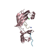
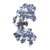
- PDB-6riu: C-terminal domain of TssA protein from T6SS of Vibrio cholerae. -
+
Open data
ID or keywords:
Loading...
-
Basic information
Entry
Database: PDB / ID: 6riu
Title
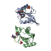
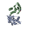
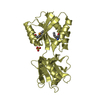
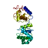
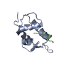
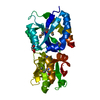
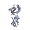
C-terminal domain of TssA protein from T6SS of Vibrio cholerae.
Components
Type VI secretion system protein TssA
Keywords
TRANSPORT PROTEIN / T6SS / Vibrio / TssA / cap
Function / homology
Type VI secretion system-associated, VCA0119 / Type VI secretion, EvfE, EvfF, ImpA, BimE, VC_A0119, VasJ / ImpA, N-terminal / ImpA, N-terminal, type VI secretion system / Type VI secretion system protein TssA / ImpA N-terminal domain-containing protein
Function and homology information
Biological species
Vibrio cholerae (bacteria)
Method
ELECTRON MICROSCOPY / single particle reconstruction / cryo EM / Resolution: 3.9 Å
Journal: EMBO J / Year: 2019 Title: Diverse roles of TssA-like proteins in the assembly of bacterial type VI secretion systems. Authors: Johannes Paul Schneider / Sergey Nazarov / Ricardo Adaixo / Martina Liuzzo / Peter David Ringel / Henning Stahlberg / Marek Basler / Abstract: Protein translocation by the bacterial type VI secretion system (T6SS) is driven by a rapid contraction of a sheath assembled around a tube with associated effectors. Here, we show that TssA-like or ...Protein translocation by the bacterial type VI secretion system (T6SS) is driven by a rapid contraction of a sheath assembled around a tube with associated effectors. Here, we show that TssA-like or TagA-like proteins with a conserved N-terminal domain and varying C-terminal domains can be grouped into at least three distinct classes based on their role in sheath assembly. The proteins of the first class increase speed and frequency of sheath assembly and form a stable dodecamer at the distal end of a polymerizing sheath. The proteins of the second class localize to the cell membrane and block sheath polymerization upon extension across the cell. This prevents excessive sheath polymerization and bending, which may result in sheath destabilization and detachment from its membrane anchor and thus result in failed secretion. The third class of these proteins localizes to the baseplate and is required for initiation of sheath assembly. Our work shows that while various proteins share a conserved N-terminal domain, their roles in T6SS biogenesis are fundamentally different.
EMPIAR-10271 (Title: Cryo electron microscopy of TssA protein from T6SS of Vibrio cholerae. Data size: 1.3 TB Data #1: Frame-averaged micrographs of TssA protein from T6SS of Vibrio cholerae [micrographs - single frame] Data #2: Aligned multi-frame micrographs of TssA protein from T6SS of Vibrio cholerae [micrographs - multiframe])
In the structure databanks used in Yorodumi, some data are registered as the other names, "COVID-19 virus" and "2019-nCoV". Here are the details of the virus and the list of structure data.
Jan 31, 2019. EMDB accession codes are about to change! (news from PDBe EMDB page)
EMDB accession codes are about to change! (news from PDBe EMDB page)
The allocation of 4 digits for EMDB accession codes will soon come to an end. Whilst these codes will remain in use, new EMDB accession codes will include an additional digit and will expand incrementally as the available range of codes is exhausted. The current 4-digit format prefixed with “EMD-” (i.e. EMD-XXXX) will advance to a 5-digit format (i.e. EMD-XXXXX), and so on. It is currently estimated that the 4-digit codes will be depleted around Spring 2019, at which point the 5-digit format will come into force.
The EM Navigator/Yorodumi systems omit the EMD- prefix.
Related info.:Q: What is EMD? / ID/Accession-code notation in Yorodumi/EM Navigator
Yorodumi is a browser for structure data from EMDB, PDB, SASBDB, etc.
This page is also the successor to EM Navigator detail page, and also detail information page/front-end page for Omokage search.
The word "yorodu" (or yorozu) is an old Japanese word meaning "ten thousand". "mi" (miru) is to see.
Related info.:EMDB / PDB / SASBDB / Comparison of 3 databanks / Yorodumi Search / Aug 31, 2016. New EM Navigator & Yorodumi / Yorodumi Papers / Jmol/JSmol / Function and homology information / Changes in new EM Navigator and Yorodumi
 Movie
Movie Controller


 Open data
 Basic information
 Components
 Keywords
 Function and homology information
 Vibrio cholerae (bacteria)
 Authors
 Switzerland, 1items
  Citation
 Structure visualization
 Downloads & links
 Other downloads
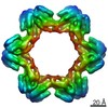

 PDBj
PDBj Assembly

 Spodoptera frugiperda (fall armyworm) / References: UniProt: A0A023PTF2, UniProt: Q9KN46*PLUS
 Sample preparation
 Electron microscopy imaging
 FIELD EMISSION GUN / Accelerating voltage: 300 kV / Illumination mode: FLOOD BEAM
 Processing
