 Movie
Movie Controller

[English] 日本語
 Yorodumi
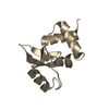
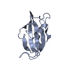
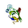
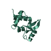
- PDB-1l8r: Structure of the Retinal Determination Protein Dachshund Reveals ... -
+ Open data

- Basic information
| Entry | Database: PDB / ID: 1l8r | ||||||
|---|---|---|---|---|---|---|---|
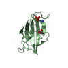
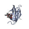
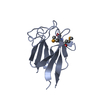
| Title | Structure of the Retinal Determination Protein Dachshund Reveals a DNA-Binding Motif | ||||||
 Components Components | Dachshund | ||||||
 Keywords Keywords | TRANSCRIPTION / winged-helix | ||||||
| Function / homology |  Function and homology information Function and homology informationdevelopment of primary female sexual characteristics / negative regulation of cell proliferation involved in contact inhibition / negative regulation of DNA biosynthetic process / regulation of nuclear cell cycle DNA replication / respiratory gaseous exchange by respiratory system / : / type B pancreatic cell proliferation / suckling behavior / negative regulation of fibroblast proliferation / negative regulation of cell migration ...development of primary female sexual characteristics / negative regulation of cell proliferation involved in contact inhibition / negative regulation of DNA biosynthetic process / regulation of nuclear cell cycle DNA replication / respiratory gaseous exchange by respiratory system / : / type B pancreatic cell proliferation / suckling behavior / negative regulation of fibroblast proliferation / negative regulation of cell migration / RNA polymerase II transcription regulatory region sequence-specific DNA binding / DNA-binding transcription repressor activity, RNA polymerase II-specific / transcription regulator complex / DNA-binding transcription factor activity, RNA polymerase II-specific / nuclear speck / RNA polymerase II cis-regulatory region sequence-specific DNA binding / negative regulation of DNA-templated transcription / regulation of transcription by RNA polymerase II / negative regulation of transcription by RNA polymerase II / Golgi apparatus / nucleoplasm / nucleus / cytosol Similarity search - Function | ||||||
| Biological species |  Homo sapiens (human) Homo sapiens (human) | ||||||
| Method |  X-RAY DIFFRACTION / SYNCHROTRON / MAD / Resolution: 1.65 Å X-RAY DIFFRACTION / SYNCHROTRON / MAD / Resolution: 1.65 Å | ||||||
 Authors Authors | Kim, S.S. / Zhang, R. / Braunstein, S.E. / Joachimiak, A. / Cvekl, A. / Hegde, R.S. | ||||||
 Citation Citation | Journal: Structure / Year: 2002 Title: Structure of the retinal determination protein Dachshund reveals a DNA binding motif. Authors: Kim, S.S. / Zhang, R.G. / Braunstein, S.E. / Joachimiak, A. / Cvekl, A. / Hegde, R.S. | ||||||
| History |
|
- Structure visualization
| Structure viewer | Molecule: MolmilJmol/JSmol |
|---|
- Downloads & links
-Download
| PDBx/mmCIF format | 1l8r.cif.gz | 52.9 KB | Display | PDBx/mmCIF format |
|---|---|---|---|---|
| PDB format | pdb1l8r.ent.gz | 37.9 KB | Display | PDB format |
| PDBx/mmJSON format | 1l8r.json.gz | Tree view | PDBx/mmJSON format | |
| Others |  Other downloads Other downloads |
-Validation report
| Arichive directory | https://data.pdbj.org/pub/pdb/validation_reports/l8/1l8rftp://data.pdbj.org/pub/pdb/validation_reports/l8/1l8r | HTTPS FTP |
|---|
-Related structure data
| Similar structure data |
|---|


-Links
 PDBj
PDBj

- Assembly
| Deposited unit | 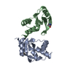
| ||||||||
|---|---|---|---|---|---|---|---|---|---|
| 1 | 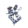
| ||||||||
| 2 | 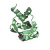
| ||||||||
| Unit cell |
|
-Components
| #1: Protein | Mass: 11285.039 Da / Num. of mol.: 2 / Fragment: DACHbox-N Source method: isolated from a genetically manipulated source Source: (gene. exp.) Homo sapiens (human) / Production host:  #2: Water | ChemComp-HOH / |  Mass: 18.015 Da / Num. of mol.: 153 / Source method: isolated from a natural source / Formula: H2O Mass: 18.015 Da / Num. of mol.: 153 / Source method: isolated from a natural source / Formula: H2OHas protein modification | Y | |
|---|
-Experimental details
-Experiment
| Experiment | Method: X-RAY DIFFRACTION / Number of used crystals: 1 |
|---|
- Sample preparation
| Crystal | Density Matthews: 2.67 Å3/Da / Density % sol: 53.88 % | |||||||||||||||||||||||||
|---|---|---|---|---|---|---|---|---|---|---|---|---|---|---|---|---|---|---|---|---|---|---|---|---|---|---|
| Crystal grow | Temperature: 277 K / Method: vapor diffusion / pH: 7.5 Details: PEG 10000, Ammonium Sulphate, pH 7.5, VAPOR DIFFUSION, temperature 277K | |||||||||||||||||||||||||
| Crystal grow | *PLUS Method: vapor diffusion, hanging drop | |||||||||||||||||||||||||
| Components of the solutions | *PLUS
|
-Data collection
| Diffraction | Mean temperature: 100 K | ||||||||||||
|---|---|---|---|---|---|---|---|---|---|---|---|---|---|
| Diffraction source | Source: SYNCHROTRON / Site: APS  / Beamline: 19-ID / Wavelength: 0.97981, 0.97904, 0.95366 / Beamline: 19-ID / Wavelength: 0.97981, 0.97904, 0.95366 | ||||||||||||
| Detector | Type: CUSTOM-MADE / Detector: CCD / Date: May 1, 2001 | ||||||||||||
| Radiation | Monochromator: Double crystal monochromator / Protocol: MAD / Monochromatic (M) / Laue (L): M / Scattering type: x-ray | ||||||||||||
| Radiation wavelength |
| ||||||||||||
| Reflection | Resolution: 1.65→25.14 Å / Num. all: 56014 / Num. obs: 53166 / % possible obs: 96.5 % / Observed criterion σ(I): 0 | ||||||||||||
| Reflection shell | Resolution: 1.65→1.71 Å / % possible all: 91.5 | ||||||||||||
| Reflection | *PLUS Lowest resolution: 50 Å / Num. obs: 26123 / Num. measured all: 227661 / Rmerge(I) obs: 0.064 | ||||||||||||
| Reflection shell | *PLUS % possible obs: 91.5 % / Rmerge(I) obs: 0.233 |
- Processing
| Software |
| |||||||||||||||||||||||||
|---|---|---|---|---|---|---|---|---|---|---|---|---|---|---|---|---|---|---|---|---|---|---|---|---|---|---|
| Refinement | Method to determine structure: MAD / Resolution: 1.65→50 Å / σ(F): 0 / Stereochemistry target values: Engh & Huber
| |||||||||||||||||||||||||
| Refinement step | Cycle: LAST / Resolution: 1.65→50 Å
| |||||||||||||||||||||||||
| Refine LS restraints |
| |||||||||||||||||||||||||
| Refinement | *PLUS Lowest resolution: 6 Å / % reflection Rfree: 9 % / Rfactor obs: 0.237 / Rfactor Rfree: 0.251 / Rfactor Rwork: 0.23 | |||||||||||||||||||||||||
| Solvent computation | *PLUS | |||||||||||||||||||||||||
| Displacement parameters | *PLUS | |||||||||||||||||||||||||
| Refine LS restraints | *PLUS
| |||||||||||||||||||||||||
| LS refinement shell | *PLUS Rfactor Rfree: 0.365 / Rfactor Rwork: 0.318 |
