 Movie
Movie Controller

[English] 日本語
 Yorodumi
- PDB-6g7c: Nt2-CTD domains of the TssA component from the type VI secretion ... -
+ Open data

- Basic information
| Entry | Database: PDB / ID: 6g7c | ||||||
|---|---|---|---|---|---|---|---|
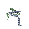
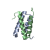
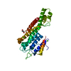
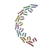
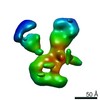
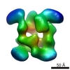
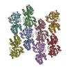
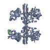
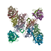
| Title | Nt2-CTD domains of the TssA component from the type VI secretion system of Aeromonas hydrophila. | ||||||
 Components Components | ImpA-related domain protein | ||||||
 Keywords Keywords | TRANSPORT PROTEIN / Type VI secretion system component / alpha helical protein / TssA / Aeromonas hydrophila | ||||||
| Function / homology | Type VI secretion system-associated, VCA0119 / Type VI secretion, EvfE, EvfF, ImpA, BimE, VC_A0119, VasJ / ImpA, N-terminal / ImpA, N-terminal, type VI secretion system / ImpA-related domain protein Function and homology information Function and homology information | ||||||
| Biological species |  Aeromonas hydrophila subsp. hydrophila ATCC 7966 (bacteria) Aeromonas hydrophila subsp. hydrophila ATCC 7966 (bacteria) | ||||||
| Method |  X-RAY DIFFRACTION / SYNCHROTRON / MOLECULAR REPLACEMENT / Resolution: 3.13 Å X-RAY DIFFRACTION / SYNCHROTRON / MOLECULAR REPLACEMENT / Resolution: 3.13 Å | ||||||
 Authors Authors | Dix, S.D. / Owen, H.J. / Sun, R. / Ahmad, A. / Shastri, S. / Spiewak, H.L. / Mosby, D.J. / Harris, M.J. / Batters, S.L. / Tzokov, S.B. ...Dix, S.D. / Owen, H.J. / Sun, R. / Ahmad, A. / Shastri, S. / Spiewak, H.L. / Mosby, D.J. / Harris, M.J. / Batters, S.L. / Tzokov, S.B. / Sedelnikova, S.E. / Baker, P.J. / Bullough, P.A. / Rice, D.W. / Thomas, M.S. | ||||||
 Citation Citation | Journal: Nat Commun / Year: 2018 Title: Structural insights into the function of type VI secretion system TssA subunits. Authors: Dix, S.R. / Owen, H.J. / Sun, R. / Ahmad, A. / Shastri, S. / Spiewak, H.L. / Mosby, D.J. / Harris, M.J. / Batters, S.L. / Brooker, T.A. / Tzokov, S.B. / Sedelnikova, S.E. / Baker, P.J. / ...Authors: Dix, S.R. / Owen, H.J. / Sun, R. / Ahmad, A. / Shastri, S. / Spiewak, H.L. / Mosby, D.J. / Harris, M.J. / Batters, S.L. / Brooker, T.A. / Tzokov, S.B. / Sedelnikova, S.E. / Baker, P.J. / Bullough, P.A. / Rice, D.W. / Thomas, M.S. | ||||||
| History |
|
- Structure visualization
| Structure viewer | Molecule: MolmilJmol/JSmol |
|---|
- Downloads & links
-Download
| PDBx/mmCIF format | 6g7c.cif.gz | 453.1 KB | Display | PDBx/mmCIF format |
|---|---|---|---|---|
| PDB format | pdb6g7c.ent.gz | 375 KB | Display | PDB format |
| PDBx/mmJSON format | 6g7c.json.gz | Tree view | PDBx/mmJSON format | |
| Others |  Other downloads Other downloads |
-Validation report
| Arichive directory | https://data.pdbj.org/pub/pdb/validation_reports/g7/6g7cftp://data.pdbj.org/pub/pdb/validation_reports/g7/6g7c | HTTPS FTP |
|---|
-Related structure data


-Links
 PDBj
PDBj- Assembly
| Deposited unit | 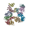
| ||||||||
|---|---|---|---|---|---|---|---|---|---|
| 1 |
| ||||||||
| Unit cell |
|
-Components
| #1: Protein | Mass: 29590.373 Da / Num. of mol.: 10 Source method: isolated from a genetically manipulated source Details: N-terminal His-tag (MGSSHHHHHHSGAP) Source: (gene. exp.) Aeromonas hydrophila subsp. hydrophila ATCC 7966 (bacteria)Gene: AHA_1844 / Production host: |
|---|
-Experimental details
-Experiment
| Experiment | Method: X-RAY DIFFRACTION / Number of used crystals: 1 |
|---|
- Sample preparation
| Crystal | Density Matthews: 3.32 Å3/Da / Density % sol: 63 % |
|---|---|
| Crystal grow | Temperature: 280 K / Method: vapor diffusion, hanging drop Details: 0.2 M sodium acetate, 0.1 M sodium citrate (pH 5.5), and 10 % (w/v) PEG 4000 |
-Data collection
| Diffraction | Mean temperature: 100 K |
|---|---|
| Diffraction source | Source: SYNCHROTRON / Site: Diamond  / Beamline: I03 / Wavelength: 0.97623 Å / Beamline: I03 / Wavelength: 0.97623 Å |
| Detector | Type: DECTRIS PILATUS 6M-F / Detector: PIXEL / Date: Mar 10, 2016 |
| Radiation | Protocol: SINGLE WAVELENGTH / Monochromatic (M) / Laue (L): M / Scattering type: x-ray |
| Radiation wavelength | Wavelength: 0.97623 Å / Relative weight: 1 |
| Reflection | Resolution: 3.13→202.08 Å / Num. obs: 67977 / % possible obs: 99.9 % / Redundancy: 3.7 % / Rmerge(I) obs: 0.113 / Rpim(I) all: 0.077 / Net I/σ(I): 5.8 |
| Reflection shell | Resolution: 3.13→3.21 Å |
- Processing
| Software |
| ||||||||||||||||||||||||||||||||||||||||||||||||||||||||||||||||||||||||||||||||||||||||||||||||||||||||||||||||||||||||||||||||||||||||||||||||||||||||||||||||||||||||||||||||||||||
|---|---|---|---|---|---|---|---|---|---|---|---|---|---|---|---|---|---|---|---|---|---|---|---|---|---|---|---|---|---|---|---|---|---|---|---|---|---|---|---|---|---|---|---|---|---|---|---|---|---|---|---|---|---|---|---|---|---|---|---|---|---|---|---|---|---|---|---|---|---|---|---|---|---|---|---|---|---|---|---|---|---|---|---|---|---|---|---|---|---|---|---|---|---|---|---|---|---|---|---|---|---|---|---|---|---|---|---|---|---|---|---|---|---|---|---|---|---|---|---|---|---|---|---|---|---|---|---|---|---|---|---|---|---|---|---|---|---|---|---|---|---|---|---|---|---|---|---|---|---|---|---|---|---|---|---|---|---|---|---|---|---|---|---|---|---|---|---|---|---|---|---|---|---|---|---|---|---|---|---|---|---|---|---|
| Refinement | Method to determine structure: MOLECULAR REPLACEMENT / Resolution: 3.13→202.08 Å / Cor.coef. Fo:Fc: 0.914 / Cor.coef. Fo:Fc free: 0.859 / SU B: 42.338 / SU ML: 0.642 / Cross valid method: THROUGHOUT / ESU R Free: 0.557 / Details: HYDROGENS HAVE BEEN ADDED IN THE RIDING POSITIONS
| ||||||||||||||||||||||||||||||||||||||||||||||||||||||||||||||||||||||||||||||||||||||||||||||||||||||||||||||||||||||||||||||||||||||||||||||||||||||||||||||||||||||||||||||||||||||
| Solvent computation | Ion probe radii: 0.8 Å / Shrinkage radii: 0.8 Å / VDW probe radii: 1.2 Å | ||||||||||||||||||||||||||||||||||||||||||||||||||||||||||||||||||||||||||||||||||||||||||||||||||||||||||||||||||||||||||||||||||||||||||||||||||||||||||||||||||||||||||||||||||||||
| Displacement parameters | Biso mean: 97.066 Å2
| ||||||||||||||||||||||||||||||||||||||||||||||||||||||||||||||||||||||||||||||||||||||||||||||||||||||||||||||||||||||||||||||||||||||||||||||||||||||||||||||||||||||||||||||||||||||
| Refinement step | Cycle: 1 / Resolution: 3.13→202.08 Å
| ||||||||||||||||||||||||||||||||||||||||||||||||||||||||||||||||||||||||||||||||||||||||||||||||||||||||||||||||||||||||||||||||||||||||||||||||||||||||||||||||||||||||||||||||||||||
| Refine LS restraints |
|