 Movie
Movie Controller


+ Open data

- Basic information
| Entry | Database: PDB / ID: 6rcu | ||||||
|---|---|---|---|---|---|---|---|
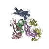
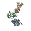
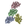
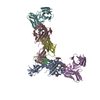
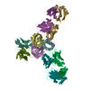
| Title | PfRH5 bound to monoclonal antibodies R5.004 and R5.016 | ||||||
 Components Components |
| ||||||
 Keywords Keywords | CELL ADHESION / Plasmodium falciparum Erythrocyte invasion Potentiating antibody Neutralising antibody Human monoclonal antibody | ||||||
| Function / homology |  Function and homology information Function and homology informationrhoptry lumen / rhoptry / symbiont entry into host / host cell membrane / bicellular tight junction / apical part of cell / heparin binding / cytoplasmic vesicle / host extracellular region / host cell surface receptor binding ...rhoptry lumen / rhoptry / symbiont entry into host / host cell membrane / bicellular tight junction / apical part of cell / heparin binding / cytoplasmic vesicle / host extracellular region / host cell surface receptor binding / symbiont entry into host cell / host cell plasma membrane / protein-containing complex / extracellular region Similarity search - Function | ||||||
| Biological species |   Homo sapiens (human) Homo sapiens (human) | ||||||
| Method |  X-RAY DIFFRACTION / SYNCHROTRON / MOLECULAR REPLACEMENT / Resolution: 4.005 Å X-RAY DIFFRACTION / SYNCHROTRON / MOLECULAR REPLACEMENT / Resolution: 4.005 Å | ||||||
 Authors Authors | Alanine, D.W.G. / Draper, S.J. / Higgins, M.K. | ||||||
| Funding support |  United Kingdom, 1items United Kingdom, 1items
| ||||||
 Citation Citation | Journal: Cell / Year: 2019 Title: Human Antibodies that Slow Erythrocyte Invasion Potentiate Malaria-Neutralizing Antibodies. Authors: Alanine, D.G.W. / Quinkert, D. / Kumarasingha, R. / Mehmood, S. / Donnellan, F.R. / Minkah, N.K. / Dadonaite, B. / Diouf, A. / Galaway, F. / Silk, S.E. / Jamwal, A. / Marshall, J.M. / Miura, ...Authors: Alanine, D.G.W. / Quinkert, D. / Kumarasingha, R. / Mehmood, S. / Donnellan, F.R. / Minkah, N.K. / Dadonaite, B. / Diouf, A. / Galaway, F. / Silk, S.E. / Jamwal, A. / Marshall, J.M. / Miura, K. / Foquet, L. / Elias, S.C. / Labbe, G.M. / Douglas, A.D. / Jin, J. / Payne, R.O. / Illingworth, J.J. / Pattinson, D.J. / Pulido, D. / Williams, B.G. / de Jongh, W.A. / Wright, G.J. / Kappe, S.H.I. / Robinson, C.V. / Long, C.A. / Crabb, B.S. / Gilson, P.R. / Higgins, M.K. / Draper, S.J. | ||||||
| History |
|
- Structure visualization
| Structure viewer | Molecule: MolmilJmol/JSmol |
|---|
- Downloads & links
-Download
| PDBx/mmCIF format | 6rcu.cif.gz | 492.8 KB | Display | PDBx/mmCIF format |
|---|---|---|---|---|
| PDB format | pdb6rcu.ent.gz | 403.4 KB | Display | PDB format |
| PDBx/mmJSON format | 6rcu.json.gz | Tree view | PDBx/mmJSON format | |
| Others |  Other downloads Other downloads |
-Validation report
| Arichive directory | https://data.pdbj.org/pub/pdb/validation_reports/rc/6rcuftp://data.pdbj.org/pub/pdb/validation_reports/rc/6rcu | HTTPS FTP |
|---|
-Related structure data


-Links
 PDBj
PDBj

- Assembly
| Deposited unit | 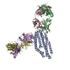
| ||||||||
|---|---|---|---|---|---|---|---|---|---|
| 1 |
| ||||||||
| Unit cell |
|
-Components
| #1: Protein | Mass: 59993.844 Da / Num. of mol.: 1 Source method: isolated from a genetically manipulated source Source: (gene. exp.) Strain: isolate 3D7 / Gene: PF3D7_0424100 / Production host:  |
|---|---|
| #2: Antibody | Mass: 24651.633 Da / Num. of mol.: 1 Source method: isolated from a genetically manipulated source Source: (gene. exp.) Homo sapiens (human) / Production host: Homo sapiens (human) |
| #3: Antibody | Mass: 23053.365 Da / Num. of mol.: 1 Source method: isolated from a genetically manipulated source Source: (gene. exp.) Homo sapiens (human) / Production host: Homo sapiens (human) |
| #4: Antibody | Mass: 23953.613 Da / Num. of mol.: 1 Source method: isolated from a genetically manipulated source Source: (gene. exp.) Homo sapiens (human) / Production host: Homo sapiens (human) |
| #5: Antibody | Mass: 50902.977 Da / Num. of mol.: 1 Source method: isolated from a genetically manipulated source Source: (gene. exp.) Homo sapiens (human) / Production host: Homo sapiens (human) |
| Has protein modification | Y |
-Experimental details
-Experiment
| Experiment | Method: X-RAY DIFFRACTION / Number of used crystals: 1 |
|---|
- Sample preparation
| Crystal | Density Matthews: 2.12 Å3/Da / Density % sol: 42 % |
|---|---|
| Crystal grow | Temperature: 298 K / Method: vapor diffusion, sitting drop / Details: 0.15 M DL malic acid, 20% PEG 3350 |
-Data collection
| Diffraction | Mean temperature: 100 K / Serial crystal experiment: N |
|---|---|
| Diffraction source | Source: SYNCHROTRON / Site: Diamond / Beamline: I04 / Wavelength: 0.9795 Å |
| Detector | Type: DECTRIS PILATUS3 6M / Detector: PIXEL / Date: Jul 31, 2017 |
| Radiation | Protocol: SINGLE WAVELENGTH / Monochromatic (M) / Laue (L): M / Scattering type: x-ray |
| Radiation wavelength | Wavelength: 0.9795 Å / Relative weight: 1 |
| Reflection | Resolution: 4.005→46.92 Å / Num. obs: 13131 / % possible obs: 99 % / Redundancy: 3.2 % / Net I/σ(I): 5.9 |
| Reflection shell | Resolution: 4.005→4.07 Å / Num. unique obs: 522 |
- Processing
| Software |
| ||||||||||||||||||||||||||||||||||||||||||
|---|---|---|---|---|---|---|---|---|---|---|---|---|---|---|---|---|---|---|---|---|---|---|---|---|---|---|---|---|---|---|---|---|---|---|---|---|---|---|---|---|---|---|---|
| Refinement | Method to determine structure: MOLECULAR REPLACEMENT / Resolution: 4.005→46.92 Å / SU ML: 0.72 / Cross valid method: FREE R-VALUE / σ(F): 1.33 / Phase error: 37.13
| ||||||||||||||||||||||||||||||||||||||||||
| Solvent computation | Shrinkage radii: 0.9 Å / VDW probe radii: 1.11 Å | ||||||||||||||||||||||||||||||||||||||||||
| Refinement step | Cycle: LAST / Resolution: 4.005→46.92 Å
| ||||||||||||||||||||||||||||||||||||||||||
| Refine LS restraints |
| ||||||||||||||||||||||||||||||||||||||||||
| LS refinement shell |
|
