 Movie
Movie Controller


+ Open data

- Basic information
| Entry | Database: PDB / ID: 5mhr | ||||||
|---|---|---|---|---|---|---|---|
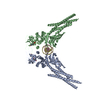
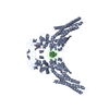
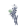
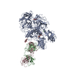
| Title | T3D reovirus sigma1 complexed with 9BG5 Fab fragments | ||||||
 Components Components |
| ||||||
 Keywords Keywords | IMMUNE SYSTEM / reovirus sigma1 / virus antibody complex / neutralization / VIRAL PROTEIN | ||||||
| Function / homology |  Function and homology information Function and homology information | ||||||
| Biological species |  Reovirus sp. Reovirus sp. | ||||||
| Method |  X-RAY DIFFRACTION / SYNCHROTRON / MOLECULAR REPLACEMENT / Resolution: 3 Å X-RAY DIFFRACTION / SYNCHROTRON / MOLECULAR REPLACEMENT / Resolution: 3 Å | ||||||
 Authors Authors | Stehle, T. / Dietrich, M.H. | ||||||
| Funding support |  United States, 1items United States, 1items
| ||||||
 Citation Citation | Journal: J. Virol. / Year: 2017 Title: Structural Insights into Reovirus sigma 1 Interactions with Two Neutralizing Antibodies. Authors: Dietrich, M.H. / Ogden, K.M. / Katen, S.P. / Reiss, K. / Sutherland, D.M. / Carnahan, R.H. / Goff, M. / Cooper, T. / Dermody, T.S. / Stehle, T. | ||||||
| History |
|
- Structure visualization
| Structure viewer | Molecule: MolmilJmol/JSmol |
|---|
- Downloads & links
-Download
| PDBx/mmCIF format | 5mhr.cif.gz | 1.1 MB | Display | PDBx/mmCIF format |
|---|---|---|---|---|
| PDB format | pdb5mhr.ent.gz | 947.9 KB | Display | PDB format |
| PDBx/mmJSON format | 5mhr.json.gz | Tree view | PDBx/mmJSON format | |
| Others |  Other downloads Other downloads |
-Validation report
| Arichive directory | https://data.pdbj.org/pub/pdb/validation_reports/mh/5mhrftp://data.pdbj.org/pub/pdb/validation_reports/mh/5mhr | HTTPS FTP |
|---|
-Related structure data
| Related structure data |  5mhsC  1figS  2oj5S S: Starting model for refinement C: citing same article ( |
|---|---|
| Similar structure data |


-Links
 PDBj
PDBj


- Assembly
| Deposited unit | 
| ||||||||
|---|---|---|---|---|---|---|---|---|---|
| 1 | 
| ||||||||
| 2 | 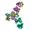
| ||||||||
| Unit cell |
|
-Components
| #1: Protein | Mass: 17921.033 Da / Num. of mol.: 6 Source method: isolated from a genetically manipulated source Source: (gene. exp.) Reovirus sp. / Gene: S1 / Production host:  #2: Antibody | Mass: 23282.822 Da / Num. of mol.: 6 / Source method: isolated from a natural source Details: DUE TO POORLY DEFINED ELECTRON DENSITY THE CONSTANT REGION OF THE FAB FRAGMENT WAS NOT BUILT FOR CHAIN J AND CHAIN L Source: (natural) #3: Antibody | Mass: 23527.369 Da / Num. of mol.: 6 / Source method: isolated from a natural source Details: DUE TO POORLY DEFINED ELECTRON DENSITY THE CONSTANT REGION OF THE FAB FRAGMENT WAS NOT BUILT FOR CHAIN K AND CHAIN M Source: (natural) Has protein modification | Y | |
|---|
-Experimental details
-Experiment
| Experiment | Method: X-RAY DIFFRACTION / Number of used crystals: 1 |
|---|
- Sample preparation
| Crystal | Density Matthews: 3.18 Å3/Da / Density % sol: 61.33 % |
|---|---|
| Crystal grow | Temperature: 277 K / Method: vapor diffusion, hanging drop / pH: 7 Details: 11% (w/v) PEG8000, 0.1 M Tris pH 7.0, 0.2 M magnesium chloride |
-Data collection
| Diffraction | Mean temperature: 100 K |
|---|---|
| Diffraction source | Source: SYNCHROTRON / Site: SLS  / Beamline: X06SA / Wavelength: 1 Å / Beamline: X06SA / Wavelength: 1 Å |
| Detector | Type: DECTRIS PILATUS 2M-F / Detector: PIXEL / Date: Mar 22, 2015 |
| Radiation | Protocol: SINGLE WAVELENGTH / Monochromatic (M) / Laue (L): M / Scattering type: x-ray |
| Radiation wavelength | Wavelength: 1 Å / Relative weight: 1 |
| Reflection | Resolution: 3→49.045 Å / Num. obs: 94667 / % possible obs: 98.5 % / Redundancy: 3.6 % / Biso Wilson estimate: 55.9 Å2 / Net I/σ(I): 13.08 |
| Reflection shell | Resolution: 3→3.08 Å / Redundancy: 3.7 % / Mean I/σ(I) obs: 1.89 / % possible all: 98.4 |
- Processing
| Software |
| |||||||||||||||||||||||||||||||||||||||||||||||||||||||||||||||||||||||||||||||||||||||||||||||||||||||||||||||||||||||||||||||||||||||||||||||||||||||||||||||||||||||||||||||||||||||||||||||||||||||||||||||||||||||||
|---|---|---|---|---|---|---|---|---|---|---|---|---|---|---|---|---|---|---|---|---|---|---|---|---|---|---|---|---|---|---|---|---|---|---|---|---|---|---|---|---|---|---|---|---|---|---|---|---|---|---|---|---|---|---|---|---|---|---|---|---|---|---|---|---|---|---|---|---|---|---|---|---|---|---|---|---|---|---|---|---|---|---|---|---|---|---|---|---|---|---|---|---|---|---|---|---|---|---|---|---|---|---|---|---|---|---|---|---|---|---|---|---|---|---|---|---|---|---|---|---|---|---|---|---|---|---|---|---|---|---|---|---|---|---|---|---|---|---|---|---|---|---|---|---|---|---|---|---|---|---|---|---|---|---|---|---|---|---|---|---|---|---|---|---|---|---|---|---|---|---|---|---|---|---|---|---|---|---|---|---|---|---|---|---|---|---|---|---|---|---|---|---|---|---|---|---|---|---|---|---|---|---|---|---|---|---|---|---|---|---|---|---|---|---|---|---|---|---|
| Refinement | Method to determine structure: MOLECULAR REPLACEMENT Starting model: 2OJ5 AND 1FIG Resolution: 3→49.05 Å / SU ML: 0.42 / Cross valid method: FREE R-VALUE / σ(F): 1.97 / Phase error: 25.66
| |||||||||||||||||||||||||||||||||||||||||||||||||||||||||||||||||||||||||||||||||||||||||||||||||||||||||||||||||||||||||||||||||||||||||||||||||||||||||||||||||||||||||||||||||||||||||||||||||||||||||||||||||||||||||
| Solvent computation | Shrinkage radii: 0.9 Å / VDW probe radii: 1.11 Å | |||||||||||||||||||||||||||||||||||||||||||||||||||||||||||||||||||||||||||||||||||||||||||||||||||||||||||||||||||||||||||||||||||||||||||||||||||||||||||||||||||||||||||||||||||||||||||||||||||||||||||||||||||||||||
| Refinement step | Cycle: LAST / Resolution: 3→49.05 Å
| |||||||||||||||||||||||||||||||||||||||||||||||||||||||||||||||||||||||||||||||||||||||||||||||||||||||||||||||||||||||||||||||||||||||||||||||||||||||||||||||||||||||||||||||||||||||||||||||||||||||||||||||||||||||||
| Refine LS restraints |
| |||||||||||||||||||||||||||||||||||||||||||||||||||||||||||||||||||||||||||||||||||||||||||||||||||||||||||||||||||||||||||||||||||||||||||||||||||||||||||||||||||||||||||||||||||||||||||||||||||||||||||||||||||||||||
| LS refinement shell |
|
