 Movie
Movie Controller

[English] 日本語
 Yorodumi
- PDB-7k66: Structure of Blood Coagulation Factor VIII in Complex with an Ant... -
+ Open data

- Basic information
| Entry | Database: PDB / ID: 7k66 | |||||||||||||||||||||
|---|---|---|---|---|---|---|---|---|---|---|---|---|---|---|---|---|---|---|---|---|---|---|
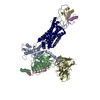
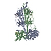
| Title | Structure of Blood Coagulation Factor VIII in Complex with an Anti-C1 Domain Pathogenic Antibody Inhibitor | |||||||||||||||||||||
 Components Components |
| |||||||||||||||||||||
 Keywords Keywords | BLOOD CLOTTING/Immune System / Antibody / inhibitor / BLOOD CLOTTING / BLOOD CLOTTING-Immune System complex | |||||||||||||||||||||
| Function / homology |  Function and homology information Function and homology informationDefective F8 accelerates dissociation of the A2 domain / Defective F8 binding to the cell membrane / Defective F8 secretion / Defective F8 sulfation at Y1699 / Gamma carboxylation, hypusinylation, hydroxylation, and arylsulfatase activation / Defective F8 binding to von Willebrand factor / blood coagulation, intrinsic pathway / platelet alpha granule / Cargo concentration in the ER / Defective factor IX causes thrombophilia ...Defective F8 accelerates dissociation of the A2 domain / Defective F8 binding to the cell membrane / Defective F8 secretion / Defective F8 sulfation at Y1699 / Gamma carboxylation, hypusinylation, hydroxylation, and arylsulfatase activation / Defective F8 binding to von Willebrand factor / blood coagulation, intrinsic pathway / platelet alpha granule / Cargo concentration in the ER / Defective factor IX causes thrombophilia / Defective cofactor function of FVIIIa variant / Defective F9 variant does not activate FX / COPII-coated ER to Golgi transport vesicle / COPII-mediated vesicle transport / Defective F8 cleavage by thrombin / : / : / endoplasmic reticulum-Golgi intermediate compartment membrane / platelet alpha granule lumen / acute-phase response / Golgi lumen / blood coagulation / Platelet degranulation / oxidoreductase activity / endoplasmic reticulum lumen / copper ion binding / : / extracellular region / plasma membrane Similarity search - Function | |||||||||||||||||||||
| Biological species |   Homo sapiens (human) Homo sapiens (human) | |||||||||||||||||||||
| Method |  X-RAY DIFFRACTION / SYNCHROTRON / MOLECULAR REPLACEMENT / Resolution: 3.92 Å X-RAY DIFFRACTION / SYNCHROTRON / MOLECULAR REPLACEMENT / Resolution: 3.92 Å | |||||||||||||||||||||
 Authors Authors | Childers, K.C. / Gish, J. / Jarvis, L. / Peters, S. / Garrels, C. / Smith, I.W. / Spencer, H.T. / Spiegel, P.C. | |||||||||||||||||||||
| Funding support |  United States, 6items United States, 6items
| |||||||||||||||||||||
 Citation Citation | Journal: Blood / Year: 2021 Title: Structure of blood coagulation factor VIII in complex with an anti-C1 domain pathogenic antibody inhibitor. Authors: Gish, J.S. / Jarvis, L. / Childers, K.C. / Peters, S.C. / Garrels, C.S. / Smith, I.W. / Spencer, H.T. / Doering, C.B. / Lollar, P. / Spiegel, P.C. | |||||||||||||||||||||
| History |
|
- Structure visualization
| Structure viewer | Molecule: MolmilJmol/JSmol |
|---|
- Downloads & links
-Download
| PDBx/mmCIF format | 7k66.cif.gz | 370.7 KB | Display | PDBx/mmCIF format |
|---|---|---|---|---|
| PDB format | pdb7k66.ent.gz | 284.4 KB | Display | PDB format |
| PDBx/mmJSON format | 7k66.json.gz | Tree view | PDBx/mmJSON format | |
| Others |  Other downloads Other downloads |
-Validation report
| Arichive directory | https://data.pdbj.org/pub/pdb/validation_reports/k6/7k66ftp://data.pdbj.org/pub/pdb/validation_reports/k6/7k66 | HTTPS FTP |
|---|
-Related structure data
| Related structure data |  6mf0S S: Starting model for refinement |
|---|---|
| Similar structure data |


-Links
 PDBj
PDBj


- Assembly
| Deposited unit | 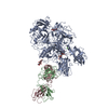
| ||||||||||||
|---|---|---|---|---|---|---|---|---|---|---|---|---|---|
| 1 |
| ||||||||||||
| Unit cell |
|
-Components
-Protein , 1 types, 1 molecules A
| #1: Protein | Mass: 168287.047 Da / Num. of mol.: 1 Source method: isolated from a genetically manipulated source Source: (gene. exp.) Homo sapiens (human)Gene: F8, CF8, F8, F8C / Production host:  Cricetulus griseus (Chinese hamster) / References: UniProt: P12263, UniProt: P00451 Cricetulus griseus (Chinese hamster) / References: UniProt: P12263, UniProt: P00451 |
|---|
-Antibody , 2 types, 2 molecules BC
| #2: Antibody | Mass: 23979.773 Da / Num. of mol.: 1 Source method: isolated from a genetically manipulated source Source: (gene. exp.) |
|---|---|
| #3: Antibody | Mass: 23297.727 Da / Num. of mol.: 1 Source method: isolated from a genetically manipulated source Source: (gene. exp.) |
-Sugars , 3 types, 3 molecules 
| #4: Polysaccharide | alpha-D-mannopyranose-(1-3)-[alpha-D-mannopyranose-(1-6)]beta-D-mannopyranose-(1-4)-2-acetamido-2- ...alpha-D-mannopyranose-(1-3)-[alpha-D-mannopyranose-(1-6)]beta-D-mannopyranose-(1-4)-2-acetamido-2-deoxy-beta-D-glucopyranose-(1-4)-2-acetamido-2-deoxy-beta-D-glucopyranose Source method: isolated from a genetically manipulated source |
|---|---|
| #5: Polysaccharide | 2-acetamido-2-deoxy-beta-D-glucopyranose-(1-4)-2-acetamido-2-deoxy-beta-D-glucopyranose Source method: isolated from a genetically manipulated source |
| #6: Sugar | ChemComp-NAG /  Type: D-saccharide, beta linking / Mass: 221.208 Da / Num. of mol.: 1 / Source method: obtained synthetically / Formula: C8H15NO6 Type: D-saccharide, beta linking / Mass: 221.208 Da / Num. of mol.: 1 / Source method: obtained synthetically / Formula: C8H15NO6 |
-Non-polymers , 4 types, 4 molecules 


| #7: Chemical | ChemComp-CU /  Mass: 63.546 Da / Num. of mol.: 1 / Source method: obtained synthetically / Formula: Cu Mass: 63.546 Da / Num. of mol.: 1 / Source method: obtained synthetically / Formula: Cu |
|---|---|
| #8: Chemical | ChemComp-CA /  Mass: 40.078 Da / Num. of mol.: 1 / Source method: obtained synthetically / Formula: Ca Mass: 40.078 Da / Num. of mol.: 1 / Source method: obtained synthetically / Formula: Ca |
| #9: Chemical | ChemComp-ZN /  Mass: 65.409 Da / Num. of mol.: 1 / Source method: obtained synthetically / Formula: Zn Mass: 65.409 Da / Num. of mol.: 1 / Source method: obtained synthetically / Formula: Zn |
| #10: Water | ChemComp-HOH / Mass: 18.015 Da / Num. of mol.: 1 / Source method: isolated from a natural source / Formula: H2O |
-Details
| Has ligand of interest | N |
|---|---|
| Has protein modification | Y |
-Experimental details
-Experiment
| Experiment | Method: X-RAY DIFFRACTION / Number of used crystals: 1 |
|---|
- Sample preparation
| Crystal | Density Matthews: 2.99 Å3/Da / Density % sol: 58.83 % |
|---|---|
| Crystal grow | Temperature: 293 K / Method: vapor diffusion, hanging drop / pH: 7.7 / Details: 17.5 %(w/v) PEG 1500, 50 mM HEPES (pH 7.7) |
-Data collection
| Diffraction | Mean temperature: 80 K / Serial crystal experiment: N |
|---|---|
| Diffraction source | Source: SYNCHROTRON / Site: ALS / Beamline: 5.0.2 / Wavelength: 1 Å |
| Detector | Type: DECTRIS PILATUS3 S 6M / Detector: PIXEL / Date: Mar 28, 2019 |
| Radiation | Protocol: SINGLE WAVELENGTH / Monochromatic (M) / Laue (L): M / Scattering type: x-ray |
| Radiation wavelength | Wavelength: 1 Å / Relative weight: 1 |
| Reflection | Resolution: 3.92→49.49 Å / Num. obs: 23431 / % possible obs: 97.61 % / Redundancy: 2 % / Biso Wilson estimate: 147.58 Å2 / CC1/2: 0.994 / CC star: 0.999 / Rmerge(I) obs: 0.09953 / Net I/σ(I): 5.74 |
| Reflection shell | Resolution: 3.92→4.06 Å / Redundancy: 2 % / Rmerge(I) obs: 0.9323 / Num. unique obs: 2201 / CC1/2: 0.342 / CC star: 0.714 / % possible all: 94.06 |
- Processing
| Software |
| |||||||||||||||||||||
|---|---|---|---|---|---|---|---|---|---|---|---|---|---|---|---|---|---|---|---|---|---|---|
| Refinement | Method to determine structure: MOLECULAR REPLACEMENT Starting model: 6MF0 Resolution: 3.92→49.49 Å / SU ML: 0.7741 / Cross valid method: FREE R-VALUE / σ(F): 1.33 / Phase error: 33.7576 Stereochemistry target values: GeoStd + Monomer Library + CDL v1.2
| |||||||||||||||||||||
| Solvent computation | Shrinkage radii: 0.9 Å / VDW probe radii: 1.11 Å / Solvent model: FLAT BULK SOLVENT MODEL | |||||||||||||||||||||
| Displacement parameters | Biso mean: 148.55 Å2 | |||||||||||||||||||||
| Refinement step | Cycle: LAST / Resolution: 3.92→49.49 Å
|
